Antipyretics for children are prescribed by a pediatrician. But there are emergency situations with fever when the child needs to be given medicine immediately. Then the parents take responsibility and use antipyretic drugs. What is allowed to be given to infants? How can you lower the temperature in older children? What medications are the safest?
The formation of the human skeleton begins with its individual parts in the womb and lasts until almost 25 years. During this time, the body grows, and the bones gradually increase in length and width.
A feature of the formation of the skeleton is the unevenness and phasing of its growth and the distal direction from top to bottom. Those parts of the musculoskeletal system that receive the maximum axial load mature faster than others. This refers to tubular bones with their articular ends where muscles are attached. Ossification nuclei are located here, which also appear as the organism develops. If this happens in a timely manner according to age, then the development process occurs normally.
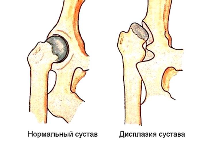
One of the most important segments is the bones of the hip joint. When the development or ossification of the nuclei is delayed, congenital hip dislocation may occur.
General understanding of kernels
Ossification nuclei are only a diagnostic sign indicating the development of the joint. There are no external signs, but the result is complete maturation of all elements of the joint.
This process has its own characteristics:
- Ossification first occurs where the first load occurs.
- Ossification nuclei in the head of the femur must appear before the child can begin to crawl and sit.
- The nuclei are first formed along its upper part (anatomically this is the roof of the joint). With its timely formation, the baby begins to stand freely on his feet, and then gradually learns to walk.
- The first ossification nuclei of the hip joints should appear precisely in the femoral head and the upper part of the acetabulum. Otherwise, the hip joint is delayed in its development and the risk of congenital dislocation in the child increases. The diagnosis becomes synonymous with hip dysplasia.
If there are no ossification nuclei in the hip joint on ultrasound, then this is called aplasia.
Statistics
Dysplasia is common in all countries (2-3%), but in different ways, depending on racial and ethnic characteristics. For example, in the United States, the likelihood of its occurrence is significantly reduced in African American children.
In the Russian Federation, in environmentally unfavorable regions, the probability of having a child with this diagnosis reaches 12%. A direct connection between the occurrence of dysplasia and tight swaddling of the baby's straightened legs has been noted.
The population of tropical countries does not swaddle newborns, they carry them on their backs, and the incidence rate here is noticeably lower.
Proof is that in Japan, for example, the tradition of tight swaddling was changed by a national project in 1975. As a result, the probability of congenital hip dislocation decreased from 3.5 to 0.2%.
The pathology most often occurs in girls (80%), a third of cases are familial diseases.
Congenital dislocation of the hip is detected many times more often with breech presentation of the fetus and toxicosis. More often the left hip joint is affected (60%), less often the right one (20%) or both (20%).
Bone nuclei of the hip joint

Bone tissue is formed in the fetus in the womb, at 3-5 months of pregnancy. Then the formation of the TBS begins. At the birth of a child, the size of the nuclei is 3-6 mm - this is the norm.
In premature babies, the ossification nuclei of the hip joints are smaller in size. But normal children can also have small nuclei. If the nuclei are missing, this is considered a pathology. If the nuclei do not appear during the first year of life, the functioning of the hip joint will not be correct.
Core pathologies
If a newborn does not have a dislocation in the pelvic area and the joint works normally, then with the slow development of the nuclei this is not considered a pathology. If irregularities and dislocation are detected, but there are no bone nuclei, then this is a pathology dangerous to health.
Normal process
There are 3 stages of normal development:
- From the formation of hip joint elements in the fetus to the first 3-4 months of life. The norm for the ossification nuclei of the hip joints in the first months of a child’s life is 3-6 mm in diameter.
- The second stage takes place between the ages of 6 months and 1.5 years. Bone nuclei develop at maximum speed, and cartilage tissue begins to gradually be replaced by bone.
- The third stage lasts until adolescence. Here the individual nuclei merge into strong plates. The lower and central parts of the acetabulum ossify.
The correct development of the ossification nuclei of the hip joints goes in parallel with the development of the child, first he learns to crawl and sit, and soon he can stand and walk.
In the fetus
Ultrasound during this period can only show gross anomalies in the development of the hip joint in the form of a complete absence of ossification nuclei or other deformations. Dysplasia is not detected on it.
In children
After birth, the newborn begins the processes of skeletal construction. And this is connected with the baby’s movements. Active leg movements develop thigh muscles. This causes blood flow to the deep parts of the bone. Dormant cells are put to work, destroyers and builders of bone beams appear. The replacement mechanism is accelerated by the fact that several bone nuclei appear.
The largest ossification nuclei are in the head of the femur, in its central sections. Simultaneously with the head of the femur, the acetabulum begins to form. It takes on its final form when the child stands on his feet. The norms of ossification nuclei, which, as already mentioned, are 3-6 mm, can be checked by ultrasound, but not earlier than in the 4th month of the baby’s life.
How to determine?

A diagnosis such as hip dysplasia is made on the basis of clinical manifestations and results of ultrasound and x-rays. These are extremely important and informative diagnostic methods, but they are secondary to the clinic.
An orthopedist should promptly suspect dysplasia while still in the maternity hospital and register the child. Such children are prescribed special treatment.
Correct formation of the joint can be determined by a number of tests:
- Skin folds on the thighs and under the buttocks are visually visible. Normally they are symmetrical.
- Hip abduction - the child's legs are bent, pressed against the stomach and then gently spread to the sides. Normally this happens easily. With dysplasia, dilation is limited - this is pre-luxation, and the tone of the thigh muscles is increased.
- At the same time, slipping is noted - when the legs are abducted from the affected side, a click is noted. This is the Ortolani-Marx symptom, and he speaks of poor fixation of the head. It is a subluxation, and the dislocation itself is determined when the child begins to walk. The baby may have a limp or have
- Shortening of one limb may occur. Even if one of these tests shows a positive result, an ultrasound scan is required.
If there is no ossification on both sides, this is not considered a serious pathology, since osteogenesis is still noted. But the unilateral process of delayed ossification nuclei requires immediate hospital treatment.
No cores
In some cases, aplasia or absence of ossification nuclei in the components of the hip joint is observed. In such cases, the body itself tries to exclude the joint from working. The violations are as follows: the legs are asymmetrical, any movements are sharply limited or impossible.
On ultrasound, ossification nuclei are absent and the components of the joint remain at the cartilaginous level. They do not contain dense inclusions and are homogeneous. The joint is deformed. The acetabulum gradually flattens and is no longer able to withstand pressure.
The femoral head emerges from the socket and its roundness disappears. The outcome is arthrosis - the joint is destroyed. The cartilage tissue becomes scarred and a bone callus occurs. Therefore, the only solution is joint replacement.
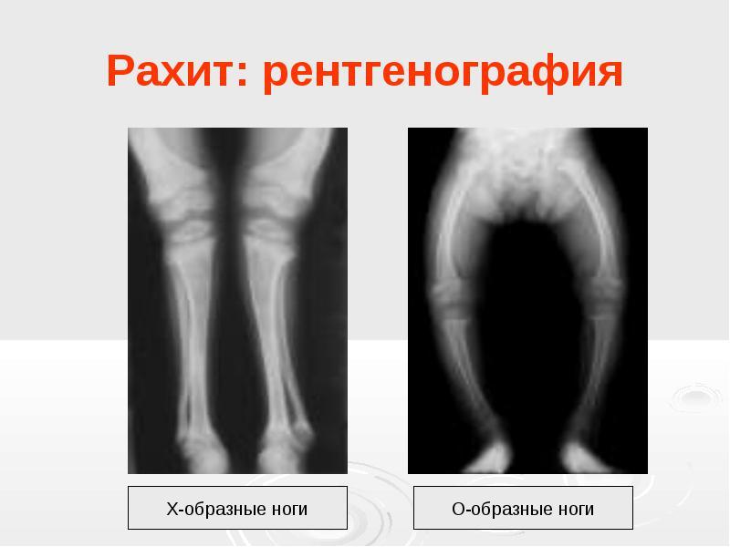
Etiology of ossification

Ossification usually occurs in 50% of rachitic patients. This is due to a lack of nutrients, B vitamins and minerals (calcium, iron, iodine, phosphorus) in the tissues of muscles, ligaments and bones. The lack of formation of ossification nuclei in children is also associated with this.
The appearance of dysplasia may be associated with malpresentation of the fetus; in children who are bottle-fed, when immunity decreases. A lot depends on the health of the mother and father: for example, the presence of diabetes, thyroid disorders, hormonal imbalances. The child's metabolism is disrupted. The reason for the absence of bone nuclei can be a twin pregnancy, gynecological pathologies in the mother in the form of uterine hypertonicity, infections and viruses during pregnancy, the mother’s age over 40 years, severe toxicosis, heredity (every fifth case), premature birth, spinal diseases in the mother, large fetus.
Development of TBS
The formation of the ossification nucleus of the femoral heads is noted at 5-6 months, and by 5-6 years the process accelerates tenfold. At 15-17 years old, cartilage is completely replaced by bone tissue. The femoral neck continues to grow until the age of 20, before the cartilage is replaced by bone.
Therapy for dysplasia
Therapy should only be prescribed by a doctor, and parents must strictly follow his recommendations. Parents need to be patient and strong, because the treatment process will be long.
The process of establishing normal development of nuclei in the hip joint area includes:
- treatment and prevention of rickets using ultraviolet irradiation and vitamin D intake;
- using a splint to realign the joint;
- electrophoresis with phosphorus and calcium, aminophylline on the lower back, procedures with bischofite;
- paraffin applications;
- massage and therapeutic exercises.
After therapy, an ultrasound is repeated to evaluate the effectiveness of treatment. During treatment, the baby should not be sat down or placed on its feet. The earlier therapy is started, the better the result will be. Exercise therapy and massage are used to strengthen and develop muscles.
It makes sense to use exercise therapy even if the child does not have dysplasia as such, but does have a genetic predisposition. Then the exercises are performed lying down, without putting stress on the joints.
Carrying out a massage

It can be carried out even with tires, without removing them. For aplasia, stroking and rubbing are indicated.
Rules for performing massage:
- the child should lie on a changing table with a flat surface;
- cover the table with a diaper, because the child may wet himself;
- the baby’s mood should be cheerful and calm;
- the child should not be hungry;
- massage is performed once a day, a course of 10-15 procedures.
There should be only 3 courses, with breaks lasting 1.5 months.
The massage complex is selected individually by a specialist. After consultation with a doctor, the mother can massage the child independently and at home. Massage is not performed if the child has:
- heat;
- ARVI;
- hernias;
- congenital heart defects.
Carrying out gymnastics

You can learn to do gymnastics yourself. The conditions are the same as for massage. Exercises are done 3-4 times during the day. Children usually love this kind of gymnastics.
Any exercise should be done very carefully. Gymnastics in the absence of ossification of the hip joint includes the following actions:
- Forming the frog pose while lying on your back. Ideally, when spreading your legs, your knees should reach the surface.
- Imitate the crawling position by turning the baby onto his stomach.
- Turn the baby over onto his back again, bending his straight legs. You need to touch the baby's head with them.
- Straight, straightened legs spread to the sides.
- Pull the straight legs towards the head and spread them to the sides.
- Place the child's legs in the lotus position, placing the left leg on top.
- Alternately bend your legs at the knees and at the pelvis.
Paraffin applications
They warm the tissues and remove toxins. For the procedure, only special processed paraffin is used. The duration of the first procedure does not exceed 1/4 hour, then the application time can be gradually increased to 30 minutes. Sea salt baths are also beneficial.
Orthopedic splints

- Koshlya splint - helps to fix the head of the femur in the center, fixes the hips in an extended state, but does not limit the movements of the pelvic joints.
- Pavlik stirrups are a fabric chest bandage that strengthens the hip ligaments. The legs do not straighten, but other movements are possible. Effective for up to a year.
- Freika splint - used for mild dysplasia up to 6 months of age. Do not use for dislocation. The splint keeps your hips at a 90 degree angle.
- When treating other types of pathology, Koshlya, Vilensky, Mirzoeva, Orlette splints, Gnevkovsky’s apparatus, and plaster are used.
- After one year, plaster casting is more often used to fix the legs. If the child is 1.5 years old and the dysplasia is not cured, surgery (according to Salter) is usually prescribed. The essence of pelvic osteotomy according to Salter is that the spatial position of the acetabulum is changed without changing its size.
Forecast
The prognosis for early treatment is good. In case of insufficient prevention, treatment will require joint replacement.
Preventative measures for the mother
A woman should eat well both during pregnancy and lactation. At 7 months, the baby’s diet should already include additional foods.
In addition to nutrition, regular walks in the fresh air, massage, exercise and hardening are of great importance. In autumn and winter, to prevent vitamin D hypovitaminosis, the child should receive it in drops. Also, preventive measures include wide swaddling of the baby so that the child can move his legs freely.
Ossification in the joints of the pelvis occurs during the first twenty years of a person’s life. Even during pregnancy, the fetus develops embryos of the ossification nucleus of the hip joints; the norm at birth is 3-6 mm.
[Hide]
Anatomical features
The rudiments of the nuclei in the articular capsule of the hip joint appear during the third to fifth months of pregnancy. Since it is during this period that human bone tissue is formed. In newborns, ossification nuclei reach three to six mm in diameter. There are cases of nuclear development only by the eighth month of pregnancy. This is why it is so important that the baby is born full term.
In three to ten percent of cases of normal development and timely delivery, the child does not have balls in the hip joint. Or they are very small. But normally, the balls can grow to the desired size by 4-6 months. Full development of the hip joint lasts up to 20 years. But by age five to six, the nuclei should be ten times larger than at birth. In the absence of this standard, there is a need to check for developmental pathologies.
Role and functions in the body
The absence of ossification nuclei of the hip joints in infants or their insufficient growth before one year can provoke problems with the development of the musculoskeletal system. The normal growth and functioning of the articulation balls influences the proper development of the pelvis as a whole. To allow the child to learn to walk, keep the torso in an even position.
Pathological state of the nuclei
Delayed formation of ossification nuclei of the hip joint or their complete absence in a newborn is in most cases a serious pathology. Which subsequently significantly affects the development of the joint. When examining a baby, the doctor looks at the state of his health, which determines in which cases the slow growth of nuclei is a pathology, and when it is the norm.
In the absence of hip dislocation, the slow growth of balls in the joint is generally not regarded as a dangerous pathology. But in case of serious disorders of the musculoskeletal system, the presence of a dislocation due to the absence of balls in the joint, it is necessary to begin treatment immediately.
Reasons for deviation from the norm
Cases where ossification nuclei appear late or their growth is delayed can be caused by a number of reasons. The basis of this pathogenesis is:
- diabetes;
- pathological metabolic disorders;
- thyrotoxicosis;
- rickets (occurs in half of newborns);
- artificial nutrition.
In most cases, insufficient development of the nuclei is accompanied by a congenital pathology such as pelvic joint dysplasia. Most often, girls are susceptible to this dislocation of the hip joint. In this case, the femoral head and the center of the nucleus do not coincide, and underdevelopment of the socket and proximal part of the femoral bone is observed.
Reasons that cause dysplasia and underdevelopment of nuclei:
- infectious lesions during pregnancy;
- hereditary factors;
- advanced maternal age;
- severe toxicosis during pregnancy;
- position of the child with the buttocks forward.
Dangerous symptoms due to underdeveloped nuclei
Dysplasia develops during pregnancy, but after birth, against the background of this pathology, dislocation of the head of the femoral bone occurs as a result of the load on the joint. It is dislocations that are a dangerous symptom of problems with the development of the pelvic nuclei.
The following types of offset exist:
- Pre-dislocation – there is a limited ability to spread the child’s legs, which were previously bent at an angle of ninety degrees. The tone of the muscle tissue of the legs is increased, there is no symmetrical arrangement of folds on the hips and buttocks.
- Subluxation is a shortening of the leg in relation to the other, a clicking sensation when the femur is abducted (the head of the bone slips into the socket of the joint).
- Dislocation - obvious disturbances during walking (tension of muscle tissue, limited functionality of the hip at the time of abduction of the leg, etc.).
When the displacement of the joint is formed, weakness of the buttocks is observed, as a result of which one limb appears shorter than the other. Children up to one year old may limp, and with bilateral lesions the baby has a duck's gait.
With bilateral pathology of nuclear development, doctors do not consider this a serious problem. The same cannot be said about unilateral joint underdevelopment.
Diagnostics
If you observe the symptoms of pelvic development disorders described above in your child, you should immediately contact an orthopedist. He examines the child, asks about the history of complaints, and the characteristics of the course of pregnancy. Next, an ultrasound examination is prescribed. It is the safest for the baby and informative. Using ultrasound, you can obtain data on the presence and size of ossification nuclei and determine their functionality.
In rare cases, the doctor prescribes an x-ray. With its help, the zones and parameters of ossification of the hip joint are more clearly visible. But x-rays produce harmful radiation on a child’s body, so it is not recommended for children, especially those under three months.
Treatment methods
After diagnosis, appropriate treatment is prescribed. An important point is that the baby is not allowed to sit or walk on his own, resting on his feet. These actions contribute to the loss of the achieved treatment results. Therefore, the task of parents is to organize a safe pastime for the child.
Therapeutic measures:
- Prevention or treatment of rickets (drink vitamin D, ultraviolet irradiation also helps).
- It is necessary to wear a special splint, with the help of which the correct arrangement of the parts of the hip joint is achieved, as well as their adequate development.
- Electrophoresis with phosphorus, calcium and bischofite in the joint area.
- Massages and exercise therapy.
- Electrophoresis procedure with euphilin on the lower back and sacrum.
- Add sea salt to the baths.
- Paraffin pads at the joint location.
- Periodic ultrasound is a diagnostic tool to study the dynamics of the disease.

If you follow all the doctor's instructions, all violations with the development of nuclei usually disappear within seven to eight months. To prevent problems, you should follow a couple of preventive measures:
- a balanced diet for pregnant and nursing mothers;
- correct diet for the child (complementary feeding is introduced from five to seven months, no later);
- massages for infants;
- walks in the open air;
- taking vitamin D during autumn, winter and spring;
- monthly pediatric examinations.
Video “Dysplasia according to Dr. Komarovsky”
In the video you will see Dr. Komarovsky’s opinion on hip dysplasia.
Medical statistics of newborn children say that congenital hip dysplasia occurs in 2–3% of cases. In 80% of them, the pathology is found in girls. Delayed formation of ossification nuclei of the hip joint begins to develop in utero. During the first year of life, the pelvic bones should stabilize and begin to develop, but this does not always happen, so pediatricians pay special attention to the formation of bone tissue in the first six months of life.
A more serious pathology is hip joint aplasia. In this case, any part of the joint is missing - the femoral head or acetabulum.
Anatomical features
The pelvic bones begin to form at the 6th week of intrauterine development and complete their growth when a person turns 19–20 years old. The most important and responsible period is the intrauterine and first year of life. Since the ligaments in infants are still weak, the hip joint is unstable. In premature babies, it is immature, since it finishes finally forming at 8–9 months of intrauterine development.
The next three months show how the joint develops:
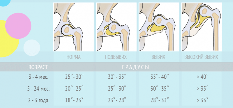
- whether the angle of the vertical position of the acetabulum decreases - normally it should decrease from 60 degrees to 50;
- whether the head of the femur coincides with the center of the round socket and the degree of insertion.
With dysplasia, ossification (the process of formation) of the hip joints in infants is delayed. The process can develop in different ways:
- in the form of fusion of the acetabulum or filling it with adipose tissue;
- increasing or decreasing the size of the femoral head.
This leads to a discrepancy between the sizes of the femoral head and socket. If the child's parents do not consult a doctor in a timely manner, the joint may completely collapse, which could lead to surgery to replace it.
Development rate by month
 At birth, the ossification nuclei of the hip joints in children measure from 3 to 6 mm, but may appear later - up to 6 months.
During the first three months, it is difficult to determine the problem because the joint is made up of cartilage, which is not visible on X-rays and ultrasound. The first information about the condition of the joints can be obtained in other ways.
At 4 months, signs of ossification of the femoral head appear. In girls, centers appear earlier than in boys. In the absence of ossification nuclei of the hip joints for 6–7 months, the process of joint formation is considered late, and doctors suggest corrective gymnastics or wearing devices that hold the baby’s legs in a bent, spread position.
If a child does not develop an ossification nucleus within 6 months, his musculoskeletal system is at risk.
Normally, with the development of the ossification nuclei of the hip joints up to 5–6 years, the growth of bone tissue should increase 10 times.
 Dysplasia is divided into degrees:
- Immaturity of the hip joint. Can be observed in healthy children. It is not a pathology.
- First degree dysplasia – preluxation. The pathology is visible on x-rays. The femur is in its place without displacement.
- Subluxation, in which the head is partially displaced, but is located in the acetabulum.
- Dislocation of the hip joint - the femoral head is separate from the socket or above it.
It has been noted that in the absence of tight swaddling with straight legs, dysplasia is capable of eliminating itself during the first six months of life.
Causes of impaired ossification of the nucleus
There are 4 groups of reasons that influence the defective formation of hip joints in children:
- Disturbances of intrauterine tissue formation. The pathology is difficult to treat, since some tissues are initially absent and cannot grow.
- Genetic predisposition. Passed on through the maternal line.
- Congenital pathologies of the spine and nervous system. Usually have concomitant musculoskeletal disorders.
- The effect of mother's hormones on the child's body. The assumption is justified, since in the first months after birth the joint begins to develop normally. Such problems are the easiest to treat, and sometimes go unnoticed.
In addition to the main reasons influencing the delay in the development of the femoral joint, there are factors that contribute to the appearance of symptoms:
- increased tone of the uterus, breech presentation of the baby, large fetus;
- insufficient intake of nutrients into the mother’s body - calcium, iodine, vitamin D, iron, vitamin E, B vitamins;
- multiple pregnancy;
- artificial feeding of a newborn;
- endocrine disorders - hypothyroidism, diabetes of one of the parents;
- having a baby in winter, when there is less sun and vitamin D is not produced in the skin, resulting in less absorption of calcium.
Viral or bacterial infections of the mother during pregnancy can provoke underdevelopment of the hip joint.
Diagnosis of pathology
 It is necessary to detect the problem earlier, since the treatment is tolerated much easier by the baby than at an older age. At the first visit to an orthopedist or traumatologist, the child is examined:
- The folds on one leg are located higher, which means there is a risk of pathology on this side. Clearly expressed asymmetry is a sign of dysplasia. Slightly expressed does not confirm the diagnosis.
- The clicking symptom is the most reliable sign by which the presence of dysplasia is determined without medical equipment. When spreading the legs and pressing on the greater trochanter, a faint crunch is heard - the femoral head is reduced into the round cavity. When mixing in the reverse order, the sound is repeated - the head comes out of the cavity.
- Normally, a baby is able to spread his legs 90 degrees. With pathology, both legs or one do not lie flat. One of the most reliable signs by which a problem is identified at the earliest stages.
- In children older than 1 year, due to untreated dysplasia, the limbs on the side of the dislocation may be shortened. To determine, place the baby on his back. The legs are bent at the hip joint. The feet are on the table. The difference is determined by the height of the knees.
Children aged 4 months and older are prescribed an x-ray or ultrasound examination.
Treatment methods

Pavlik stirrups
Children under 6 months of age are recommended to wear stirrups without limiting joint mobility. After 6 months, if there is no progress in the formation of nucleoli, a fixation structure is needed - a crossbar between the separated legs. If the development of hip joints is delayed, calcium supplements, walks in the air, and sunbathing are additionally prescribed. If the child is breastfed, calcium supplements are prescribed to the mother.
Massage
Massage begins from the first days of life if examination reveals a delay in the development of the hip joint. With regular massage procedures, the pathology can disappear on its own by the age of three months.
Gymnastics
Physical therapy is also done early. This improves blood supply to the joints and helps strengthen muscles and ligaments. Exercises are done in two positions: lying on your back and on your stomach. During the treatment period, the child should not be seated or stood on his feet.
Paraffin applications
The temperature of the molten paraffin should be between 40 and 45 degrees for small children. The procedure is aimed at accelerating blood flow in the affected area. Muscle tissue develops better with thermal stimulation. Ozokerite is sometimes added to paraffin. To treat hip dysplasia, children are given a layer of paraffin from the buttocks to the foot in the shape of a boot. For newborns, the substance is kept on the body for 7 minutes. After 6 months – 10 minutes. After the procedure, a massage is performed. 20 paraffin wraps are recommended.
Prevention of pathology in children
Prevention begins with the mother's nutrition during pregnancy. If joint diseases have occurred in the maternal family, it can be predicted that the newborn child may have similar problems. Especially if the child is a girl.
 The baby should be given complementary foods and vitamins on time if treatment is being carried out. In autumn and winter, vitamin D is additionally prescribed. Hardening and contrast procedures on the pelvic area help.
Timely early examination of the child - at 1 month, 3 months from birth. Tight swaddling can negatively affect the condition of the joints. At the end of the last century, Japan had a program that did not recommend swaddling infants. They tried to convey information to grandmothers caring for babies so that they would not use old methods. As a result, the incidence of dysplasia in the country dropped to 0.1%.
The formation of the human skeleton normally extends over a long period of time - its individual elements mature up to the age of 25 years. This feature of the processes is due to the general growth of the body, which allows the bones to freely increase in length and width. If ossification was completed in childhood, then serious skeletal anomalies could be observed, leading to disruption or complete loss of its functions.
Due to the unevenness of growth processes, those parts of the musculoskeletal system that are subject to the greatest axial load mature faster. These include most tubular bones, especially in the area of the articular ends and areas of muscle attachment. These segments contain ossification nuclei, the timely appearance of which indicates the normal development of these formations.
These nuclei are of greatest interest in the area of the hip joint - their role in the mechanisms of occurrence has already been proven. With the help of timely diagnostics - ultrasound examination - the structure of the joint is assessed in children. And the combination of ultrasound results and even the slightest clinical signs of damage to the hip joints makes it possible to begin treatment that prevents the development of irreversible changes.
Concept
Ossification nuclei are a purely diagnostic sign that characterizes normal or pathological development of the hip joint. External signs of this process are invisible, but the result is always obvious - the complete maturation of all elements of the articulation. This occurs due to the implementation of the following mechanisms:
- Ossification of all elements of the musculoskeletal system does not occur simultaneously - initially only those parts that will take on the first load are strengthened.
- The hip joint in children up to about 6 months of age is practically not involved in any significant movements. Therefore, for crawling and sitting, at least the formation of bone nuclei in the head of the femur is necessary in order to achieve minimal mobility.
- Ossification nuclei in the acetabulum of the pelvis appear first only in the upper part, which in anatomy is called the roof. If it is formed on time, the baby will be able to stand on his feet and walk calmly.
- Therefore, initially these bone nuclei should appear in large numbers precisely in the femoral head and the upper part of the acetabulum. A decrease in their number leads to a delay in the development of the joint, which becomes a risk of developing congenital dislocation in children.
If ossification nuclei are not detected at all in the hip joint during ultrasound, then this condition is called aplasia (absence).
Normal process
Since the formation of nuclei is a physiological mechanism, it normally occurs unnoticed by the child himself and his environment. Unlike teething, the growth of bone tissue is not accompanied by any unpleasant sensations. The result of all processes is partial maturation of the hip joint, preparing it for further loads. The following three stages of normal development are distinguished:
- The first period lasts from the formation of articulation elements in the fetus until the first months of the child’s life. During it, the anatomical structures consist only of cartilage tissue, and their shape is significantly different from the structure of the hip joint in an adult.
- The second stage is the most important - it starts from about 6 months and ends by one and a half years of the baby’s life. It is at this time that the maximum development of bone nuclei is observed, which gradually replace cartilage tissue.
- The third period lasts until puberty - during it, the fusion of all individual nuclei into strong plates occurs. The last thing that normally occurs is ossification of the lower and central parts of the acetabulum.
The correct development of the nuclei is combined with stages of increasing the child’s activity - at first he learns only to crawl and sit, and soon he is able to easily stand and walk.
In the fetus

Considering the possibilities for early diagnosis of many diseases at the present time, parents of an unborn child often want to know the risk of developing congenital hip dislocation. They assume that screening (ultrasound) during pregnancy will give them this information. But in a fetus such diagnostics will be useless for the following reasons:
- The formation of the hip joint can be assessed in late pregnancy, when all parts of the fetus’s body are clearly visible.
- The detection of large bone nuclei is not a physiological process - by the time of birth in children, the articulation is formed only by connective and cartilaginous tissue.
- The joint in the fetus and newborn does not play a significant role in movements, so the first signs of maturation are observed only in the interval from 3 to 4 months.
An ultrasound examination during pregnancy will show only gross anomalies in the development of the hip joint - its complete absence or serious congenital deformities.
In children

Immediately after birth, construction processes begin in the baby’s body, allowing all organs and systems to be prepared for new conditions. First of all, they concern the musculoskeletal system, the elements of which begin to grow under the influence of movements. Therefore, the formation of nuclei begins due to the following factors:
- With active movements of the legs, the thigh muscles actively contract, which increases blood flow in the deep parts of the bone.
- Normal mobility promotes the launch of dormant cells - some begin to destroy cartilage tissue, while others form bone beams in its place.
- Usually several nuclei are formed, which allows the replacement mechanisms to be accelerated. The largest formations are determined in the central parts of the femoral head, from where they spread to the surface.
- The acetabulum begins to form simultaneously with the femoral head, but acquires its final shape only after the start of standing and walking.
- For ossification to become noticeable, a certain time must pass - on ultrasound, signs of nuclei normally become noticeable between 4 and 6 months after birth.
Normal development of the hip joint is always accompanied by a natural course of developmental periods - children learn to stand and walk in time.
How to determine?

The correct formation of the articulation can be assessed without resorting to special diagnostic methods. For this purpose, a number of special tests are carried out in medicine:
- Externally, the symmetry of the skin folds on the thigh and under the buttocks is assessed. If they are at different levels, then this indicates a delay in the development of the joint.
- A hip abduction test is performed - the child’s legs are bent and pressed to the stomach, after which they are gently moved apart. Normally, in children, due to the small size of the articular cavity, they can be easily moved apart. In case of pathology of the development of the hip joint, dilution is limited.
- Simultaneously with the previous test, slipping is assessed - if a click is felt when the legs are abducted on one or the other side. Slowing down of ossification contributes to such a crunch, which is caused by poor fixation of the femoral head in the unformed cavity.
Such tests can be easily carried out at home, taking precautions so as not to injure the child. If at least one of them is positive, then it is necessary to evaluate the ossification nuclei.
Pathological variants

Violation of the physiological mechanisms of bone tissue formation in the elements of the hip joint primarily affects the development of the child. When the muscles and soft tissues are fully formed, the time comes for the implementation of the supporting and motor functions of the joint. But anatomically he is not ready for such work, which causes his gradual deformation.
Such changes are preceded by anomalies in the formation of ossification nuclei in the femoral head and acetabulum. They are based on the following changes in the child’s body:
- Most often, the mechanism is disrupted already in the womb, which leads to errors in the formation of cells that are the source of bone tissue. This is facilitated by various endocrine diseases, infections or intoxications during pregnancy.
- Currently, the incidence of congenital hip dislocations caused by rickets in children has decreased. But this problem still remains relevant, since a deficiency of vitamin D and calcium in children leads to various skeletal lesions (including hip joints).
- Another common option is the birth of a premature baby. Due to the immaturity of all organs and systems, such children often experience various developmental deviations.
Absolutely all infants do not undergo an ultrasound of the hip joints - it is performed only according to indications based on external examination data.
Slowdown

The delay in the formation of foci of ossification in the femoral head and glenoid cavity until the baby’s first steps rarely becomes noticeable. A slowdown in the formation of nuclei in the hip joint is accompanied by the following symptoms:
- Externally, changes may not be noticeable for up to a year - the symmetry of the skin folds is maintained, hip abduction is slightly limited.
- The main manifestations become noticeable only on ultrasound - at the age of about 6 months, the bone nuclei are small.
- When observed over time, their gradual increase occurs, which still lags behind the rate of growth and development of the child.
- Over time, a gradual enlargement of the nuclei should be observed, as well as their partial fusion. When you slow down, by the time you start walking, only a partial connection is determined in the central sections.
Since the processes of bone tissue formation still occur, the outcome of delayed ossification without treatment is usually congenital subluxation of the hip.
Absence
In some cases, complete aplasia of the bone nuclei in the components of the hip joint is observed. A serious anomaly becomes an obstacle to the development of its supporting and functional qualities, after which the body tries to turn off the defective connection from working. Aplasia is accompanied by the following disorders:
- Even without the supporting function, damage to the joint becomes noticeable - externally the legs lose symmetry, any movements are difficult or impossible.
- By the time the first nuclei appear on ultrasound, their signs are not revealed - the glenoid cavity and the femoral head are formed only from cartilage tissue. They have a characteristic homogeneous appearance (without additional dense inclusions).
- When observing dynamics, signs of ossification are not detected - gradually the joint begins to lose its original configuration, deforming under the influence of muscles and ligaments.
- The acetabulum gradually flattens, since its roof, due to its softness, is not able to withstand constant pressure. The head of the femur moves higher, after which it begins to gradually lose its rounded shape.
The outcome of this type of disorder is always that an overly soft joint cannot withstand the load, which leads to its gradual destruction. Due to regular damage, cartilage tissue is replaced with a scar, which gradually takes on the appearance of a callus. Therefore, the only option for help in this case is joint replacement.
On TV
Dr. Komarovsky dedicated one of his programs to the topic of development of the hip joint, where he explained in diagrams and pictures the mechanisms of development of ossification nuclei in normal and pathological conditions. In the program he addressed the following issues:
- Physiological processes occurring in the hip joint during the growth and development of a child.
- Reasons that influence the proper formation of the musculoskeletal system in children, as well as unfavorable factors that slow down growth mechanisms.
- Methods of diagnosis and treatment for delayed formation of bone nuclei, as well as timely prevention to prevent the formation of congenital hip dislocation.
The program will not only allow you to learn theoretical material about the physiological processes of growth, but will also provide a visual representation of them. In this form, it will be much easier for parents to understand what is required to give birth and raise a healthy child.


