Antipyretics for children are prescribed by a pediatrician. But there are emergency situations for fever when the child needs to be given medicine immediately. Then the parents take responsibility and use antipyretic drugs. What is allowed to give to infants? How can you bring down the temperature in older children? What medicines are the safest?
/ Canine interdigital furunculosis (canine interdigital cysts)
Canine interdigital furunculosis (canine interdigital cysts)
Text of the article and photo from the book SMALL ANIMAL DERMATOLOGY A COLOR ATLAS AND THERAPEUTIC GUIDE 2017
Translation from English: veterinarian Vasiliev AB
Peculiarities
Canine interdigital furunculosis appears as single or multiple erythematous papules; dense or fluctuating nodules; or bullae (called "cysts") on one or more paws, which are usually located in the interdigital zones. Lesions may be painful or itchy, may be ulcerated, may develop fistulas with serous hemorrhagic or purulent exudates, and may become fibrotic in the long run. Lesions may spontaneously disappear, intensify, fade, or persist indefinitely. Regional lymphadenopathy is observed frequently, but systemic symptoms of the disease are not noted. Secondary fungal and bacterial infections are often observed.
Interdigital cysts in dogs are a common problem in short-haired dog breeds. Their severity and recurrence are often exacerbated by the underlying pruritic disease type. Although the cause of the disease is unknown, the short hairs that emerge from the damaged follicle, creating a sterile boil that subsequently becomes infected, seem to be an important component of the disease. Ingrown hairs are a key feature in the development of interdigital cysts in dogs.
Differential diagnoses
Diagnosis
1 Based on medical history, clinical findings; exclusion of other differential diagnoses
2 Cytology (aspirate from nodules or unruptured interdigital cysts): present (pyo)granulomatous inflammation with no microorganism unless there is secondary infection.
3 Dermatohistopathology: multifocal, nodular to diffuse, (pyo)granulomatous dermatitis. Special staining does not detect infectious agents unless there is a secondary infection.
4 Microbial culture (biopsy samples): no bacteria, mycobacteria and fungi.
Treatment and prognosis
1 The clinician should be confident that the underlying cause of interdigital furunculosis (eg, damp environment, dirty kennel, rubbing in shorthaired breeds) is identified and corrected.
2 If fistulous lesions are secondarily infected, appropriate antibiotics or antifungals should be given for a minimum of 4–6 weeks.
3 For single lesions, surgical excision or laser ablation may be used.
4 Cleansing wipes (baby wipes, chlorhexidine-containing tampons, or other antimicrobial wipes) used every 12 to 72 hours work very well. For interdigital cysts, surgical removal of ruptured hair follicles and "ingrown" hairs with needle biopsy or laser speeds recovery. When interdigital cysts develop, topical treatment with dimethyl sulfoxide (DMSO) in combination with enrofloxacin (make a 10 mg/ml solution) and steroids (dexamethasone as a 0.1/mg/ml solution) should be used every 12 to 72 hours until the lesions resolve. To prevent recurrence, the paw should be rubbed or scraped in the direction of hair growth to remove any "ingrown" hairs.
5 Alternatively, a combination of tetracycline and niacinamide may be effective in some dogs. A positive response should be observed within 6 weeks after the start of treatment. Administer 500 mg of each drug (dogs >10 kg) or 250 mg of each drug (dogs £10 kg) orally every 8 hours until lesions improve (approximately 2–3 months). Then give each drug every 12 hours for 4-6 weeks, then try to reduce the frequency to 1 time per day for a maintenance effect. Rare reports suggest doxycycline 10 mg/kg every 12 hours until beneficial, then tapered to the lowest effective dose (doxycycline may be substituted with tetracycline).
6 Rare reports indicate that treatment at a dose of 5 mg/kg orally every 24 hours may be effective in some dogs. Once clinical improvement is achieved (usually within 6 weeks), the dose of ciclosporin should be gradually reduced to the lowest possible dose daily or every other day to maintain remission. The addition of ketoconazole (5-11 mg/kg/day orally with food) to ciclosporin may allow a reduction in the ciclosporin dose used.
7 For severe, non-surgical or multiple lesions, treatment with glucocorticosteroids may be effective. Prednisone or prednisone 2-4 mg/kg orally should be given every 24 hours. Significant improvement should be seen within 1-2 weeks. After improvement in the condition (after approximately 2-3 weeks), the dose of steroids should be gradually reduced to the lowest dose every other day, maintaining remission. In some dogs, steroid therapy may eventually be discontinued. Secondary infections are common and must be treated aggressively.
8 The prognosis is good to cautious. Lifelong drug therapy may be required to maintain remission, and interdigital fibrosis may be a permanent consequence of chronic cases.
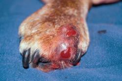
Photo 1 Interdigital cyst. A large, soft cyst in the interdigital space is typical of this condition.

Photo 2 Interdigital furunculosis of dogs.Severe swelling of the tissues in the interdigital space was caused by traumatic furunculosis and subsequent bacterial infection.

Photo 3 Interdigital cyst of dogs. Interdigital cyst with moist exudate and bruising in the surrounding tissue

Photo 4 Interdigital furunculosis of dogs. The fingers are spread apart, showing the interdigital space that looks like a bruise. The skin appears thin, with a focal area of exudate in the form of a focal abscess.

Photo 5 Interdigital furunculosis of dogs. The clinician gently squeezes the lateral parts of the lesion to demonstrate the presence of hair in the abscess cavity. This technique is not recommended as internal damage to the lesion can exacerbate cellulite and scarring.

Photo 6 Interdigital furunculosis of dogs. The extruded material includes exudate and numerous hairs. This hair acts as a foreign body and a source of recurrent secondary infection.

Photo 7 Interdigital cyst of dogs. Small interdigital cyst.

Photo 8 Interdigital furunculosis of dogs. Interdigital tissue affected due to severe pyogranulomatous infiltration leading to cellulitis

Photo 9 Interdigital furunculosis of dogs. Pronounced increase in interdigital space caused by chronic inflammation

Photo 10 Interdigital cyst of dogs. Focal interdigital cyst that is ruptured and exudes purulent exudate.

Photo 11 Interdigital furunculosis of dogs.. Pronounced interdigital cellulitis with deep ulceration.
.jpg)
Photo 12Interdigital cyst in dogs.. This interdigital cyst (interdigital furunculosis) was discovered only after that. how the fingers were spread apart to examine the interdigital space
TUMORS
Tumor cell growth, starting from a local focus, acquires new, pathological properties. These properties of cells are transferred to the next generation of cells, and thus a new type of them arises, which is the basis of the pathological process and tumor disease.
The main features of tumors are the atypical structure of their cells and tissues and unlimited growth, which continues even after the elimination of the main causes that caused their appearance. Such features are inherent in all types of tumors.
Adenocarcinomas and other malignant mixed tumors often undergo ulceration as a result of the appearance of necrotic processes due to a lack of nutrition of the tumor tissue. Most often, such processes occur as a result of trauma to the tumor. Sometimes a cyst formed in close proximity to the surface of the tumor opens and becomes infected, which causes a purulent-necrotic process. The general condition of the animal and appetite, as a rule, are satisfactory.
Tumors, or neoplasms, are pathological growths of body tissues that occur as a result of the reproduction of cellular elements under the influence of exogenous and endogenous factors.
Bone tumors. Osteosarcomas are noted in the shoulder, hip, etc. This malignant tumor is characterized by a rapid course and causes high mortality. It is mainly installed in large breeds of dogs (Boxers, Great Danes, East European Shepherds, etc.). Tumor growth occurs in the metaphyseal region, mainly the humerus and tibia. Benign bone tumors are rare in dogs.
Lameness associated with pain, limited mobility in the joint, swelling of the limb, swelling in the pathological zone are pronounced.
Practically, surgical treatment of bone sarcoma is still ineffective. Treatment should be complex with the use of chemotherapy and radiation methods. It is important to identify sick animals at an early stage of the disease.
Tumors of the upper respiratory tract. The most common neoplasms of the nasal and adnexal cavities.
Tumors of the nasal and adnexal cavities in dogs are characterized by unilateral nasal leakage, difficulty breathing, sniffling, and wheezing. The discharge is mucous or bloody. In advanced cases, deformation of the front of the head, shakiness of the teeth, displacement or loss of them are noted. Exophthalmus can be observed.
Early diagnosis and urgent surgery provide the greatest effect. Operative access - from the side of the nasal bone, which is cut longitudinally, and then the tumor is removed. Thoroughly pack the cavity. After the operation, antibiotic therapy is prescribed.
Papillomas of the mucous membrane of the buccal surface, lips, gums often occur in dogs at a young age. They have a soft, friable texture, abundantly supplied with blood vessels and therefore are easily injured and bleed. Papillomas are also located on the skin, but they are denser in consistency, painless, and keratinized in appearance.
Papillomas are removed by an operative method, if intravenous administration of a 0.5% solution of novocaine, 5 ml, after 3 days does not help (the course consists of three to four injections).
Fibrosarcomas are observed on the mucous membrane of the cheeks and gums. They are of a dense consistency, with a smooth or nodular surface, sometimes they are ulcerated -
Treatment is surgical only. After removal, they can quickly recur.
Tumors of the mammary gland. In dogs, benign and malignant neoplasms of the mammary gland are noted. A single cause of the occurrence of mammary tumors has not been established. We can only talk about some factors that are somehow related to one degree or another.
Often there are cases when dog owners, on their own conviction and on the advice of insufficiently competent persons, prefer conservative treatment, making erroneous conclusions about the nature of the disease. In such cases, time is usually lost, the tumor progresses in growth. In the presence of metastases in regional lymph nodes, surgical removal of malignant neoplasms in the mammary gland becomes useless
Sometimes the refusal of surgical treatment is motivated by the age of the animal. We do not advise dog owners to refuse surgical treatment, even if the dog is old. The progressive increase in the tumor aggravates the condition of the sick animal. There are ulcerations of the tumor due to injury or decay of its tissue, which is accompanied by purulent secretions of a putrefactive odor. It is difficult to keep such a dog. Removal of such a tumor relieves the animal of severity in cases where the tumor reaches a large size, and greatly improves the general condition of the dog. Often such animals delight the owner for a long time.
CYST
Cysts - a closed cavity formed in the body as a result of various pathological processes, having a wall and filled with contents
Depending on the mechanism of development, structural features and localization, cysts are divided into retention, tumor and cysts arising from a violation of normal development. The size, structure of the wall and content of cysts are varied, which is associated with the cause and nature of their development, as well as with localization.
Retention cysts develop in various glandular organs with a delay or complete cessation of the outflow of secretions. The latter, accumulating in the excretory duct or glandular lobule, stretches them, resulting in the formation of cavities filled with secretion. In dogs, such cysts are often found in the salivary glands.
A number of diseases in dogs at first glance do not cause unrest among the owners, as they seem quite harmless, but the consequences can be irreparable. These diseases include pathological formations. A cyst in dogs in most cases is not dangerous, but some of its types can deprive a pet of health and even lead to death.
Photo of a cyst in a dog
In some cases, a cyst can degenerate into a malignant neoplasm, so a veterinarian's diagnosis and regular monitoring are necessary.
The type of cyst is determined by many signs - the place of formation, the structural features of the cavity, the tendency to growth and development, and others.
An epidermal or cutaneous cyst is quite common. Its distinctive features include the following:
- Location - the upper layers of the epidermis.
- A cavity in the form of a circle or a sphere.
- The softness of the formation, you can check this during probing.
- The cyst does not bring pain to the dog.
- The size of the formation rarely exceeds 50 mm.
The causes of cysts formed on the surface of the skin are very diverse. Often they appear against the background of a clogged pore, which does not allow the skin secret to go out. As a result, a cavity is formed, which is gradually filled.
The pores can be clogged due to adverse external conditions - chemical emissions into the atmosphere, excessive air pollution, ozone holes. All this disrupts skin metabolism, as a result of which an external cyst may appear.
Not the last role is played by the hereditary factor. Scientists have found that the chance of an epidermal cyst in a dog is higher if its closest relatives suffered from this disease. The formation of such an etiology may be congenital. External cysts in dogs are most often found on the gums, between the toes, in the auricles.

In addition, in dogs there are cystic cavities formed on the internal organs. Most often they occur on the ovaries and mammary glands. Internal formations can disrupt the functioning of the dog's organs and systems, some types of them lead to infertility, and in case of ruptures, there is a high probability of death.
The causes of cysts are still poorly understood by modern medicine.
Symptoms of the disease
1. Cyst located on the surface of the skin
A cyst located on the surface of the skin is determined visually. Education on the gums can be detected at the initial stages. The tissues, at the site of the appearance of the cyst, swell, differ in a bright red tint. Over time, the tumor may spread throughout the gum.
During this period, quite often the dog feels unwell, quickly gets tired, maybe in some cases it rises. When examining the mouth, a white or yellow neoplasm filled with pus will be revealed. If you do not help the pet, then he will suffer from pain, asymmetry of the mouth, flux.
2. Sweat gland cyst
Sweat gland cyst - blue, blue or grayish, its size rarely exceeds 1 cm. Often a sweat gland cyst occurs in the ear cavity, in most cases it is not dangerous, the pet does not experience discomfort.
3. Follicular cyst
The follicular cyst occurs at the root of the hair, its filling is dead cells. The characteristic color is gray, the size varies from 1 mm to several cm.
4. Dermoid cyst
Dermoid cyst is a congenital malformation. It is a closed soft cavity of a rounded shape, towering above the hairline. In some cases, there is a risk of infectious damage to the nervous system, so veterinarians recommend timely treatment.
5. Cyst in the sublingual zone
The cyst may also appear in the sublingual zone. It looks like a round purulent bag of white-pink color. When a cyst forms in this place, the dog often refuses to eat, as it feels uncomfortable in the process of eating food. If the size of the formation reaches a critical size, the dog may even stop drinking, and begin to wither from exhaustion.
6. Interdigital cyst
The interdigital cyst in most cases does not cause pain in the pet, it looks like a rounded swelling. The formation is soft, the temperature in the area of the cyst is not elevated, there are both single and multiple swellings. This disease is especially susceptible. The basis of the disease is the ingrown hair into the skin.
Skin cysts have the same symptoms:
- Slowly increase in size.
- They do not cause pain, the dog behaves calmly while probing the cavity, does not try to drive the owner away from himself.
- During the probing of the cyst, its smooth structure, heterogeneity, granularity is revealed - a dangerous "bell".
- The pet gets tired quickly, sleeps more than usual, does not want to walk for a long time.
- In some cases, the body temperature rises.
- Loss of appetite.
If a seal is found on the surface of the skin or mucous membranes, an urgent need to consult a doctor to exclude oncological formations. The veterinarian will diagnose and, if necessary, prescribe treatment.
Most internal cysts are asymptomatic and are diagnosed by accident. However, sometimes the following symptoms occur:
- Increased body temperature in a pet.
- Loss of appetite.
- Weakness, unwillingness to walk, apathy.
The asymptomatic course of the disease is dangerous because the cyst may burst. In this case, the following symptoms are recorded:
- Seizures.
- Bloody discharge as a sign of internal bleeding.
- Attacks of pain during which the dog whines, behaves unnaturally, and may lose consciousness.
- Increased body temperature.
These symptoms indicate that the dog needs urgent medical attention, and it is impossible to postpone a visit to the doctor even for a few hours.
Proliferative lesions of the oral cavity are observed in dogs and cats quite often. The examination should include a complete physical examination, imaging studies, and a histopathological examination of a sufficiently good quality biopsy. Proliferative lesions are divided into reactive and neoplastic. Some of them may represent an epulis - a tumor-like growth on the gum. The most common reactive gum disease is gum hyperplasia.
Tumor lesions include odontogenic and non-odontogenic tumors. The most common odontogenic tumors are peripheral odontogenic fibroma and acantomatous adamantinoma (acantomatous ameloblastoma). The most common non-odontogenic neoplasms are malignant melanoma and squamous cell carcinoma.
The article discusses the prevalence, clinical presentation, and treatment options for proliferative lesions; special attention is paid to new methods of treatment. For most proliferative lesions, surgery remains the most important component of the treatment plan.
Proliferative lesions of the oral cavity, epulis, reactive lesions, odontogenic tumors, non-odontogenic tumors.
Introduction
Oral tumors account for approximately 5–10% of all tumors in dogs and cats. In dogs, a significant proportion of proliferative lesions are reactive or benign, while in cats, most proliferative lesions are malignant.
Proliferative lesions or local edema in the oral cavity can manifest a variety of clinical conditions, including infectious diseases. In addition, a non-healing ulcer that looks like an infection may well be malignant. The precise nature of any lesion can only be determined by histopathological examination.
Biopsy is indicated for all proliferative or other suspicious lesions such as non-healing ulcers. The main method of treatment of malignant neoplasms of the oral cavity is to carry out, if possible, a radical operation.
Clinical manifestations
Unfortunately, most owners are not accustomed to regularly inspecting the oral cavity of their animals. Thus, when contacting a doctor in most patients, the disease is already at a late stage.
Clinical manifestations typically include halitosis, tooth mobility, exfoliation of tooth enamel, bleeding from the mouth, increased salivation; with damage to the upper jaw - discharge from the nose. There are no obvious signs of pain in most patients, except in cases of tongue involvement or advanced stages of the tumor, when it interferes with chewing or leads to pathological fractures. Sometimes the main reason for contacting a veterinarian is a pronounced deformation of the muzzle of the animal.
Clinical examination
1. Direct examination
It is necessary to find out the clinical manifestations observed by the owner, the duration and progression of the lesion, previous treatment and its results. A complete physical examination should be performed to detect distant metastases.
On examination and palpation of the head, asymmetry, increased pressure in the retrobulbar region (with distal lesions of the maxillary sinuses), bleeding from the mouth or nose, and bad breath can be detected. Volumetric lesions should be carefully examined and palpated, noting the location, size and consistency of the lesion, color (abnormal pigmentation or loss of pigmentation), presence of ulcers and/or necrosis, fixation to underlying tissues, displacement of teeth, any evidence of abnormal tooth mobility, change in bone contour. An example of a survey is shown in Fig. 1.

Rice. 1. Proliferative lesion in a Cocker Spaniel. In the right half of the lower jaw, a lesion 4 cm wide, dense, of normal pigmentation, ulcerated due to trauma by opposing teeth, fixed to the underlying bone, is revealed. The teeth are displaced, but not mobile.
Regional lymph nodes should be palpated and evaluated for size, shape, and consistency, as well as possible fixation to surrounding tissues.
2. Visualization methods
Radiographic control of the state of the affected jaw is mandatory. In most cases, it is best visualized with screenless dental x-rays and intraoral x-rays.
Bone infiltration can be diagnosed by identifying differences in the severity of resorption and / or the formation of new bone tissue. Bone resorption with the standard technique is visualized only when about half of the bone mineral content has been lost. In some malignant tumors, signs of resorption of the roots of the teeth may also be detected. Common radiological signs are shown in Table 1.
|
Benign lesions |
Malignant/ aggressive lesions |
|
well-defined boundaries |
Boundaries are inaccurate or not defined |
|
Extension or thinning cortical bone |
Destruction of adjacent cortical bone |
|
Periosteal reaction: absent or smooth |
Periosteal response uneven |
|
Density: variable, often increased |
Density: variable, often reduced |
|
Teeth may be misaligned |
Floating teeth, root resorption possible |
Table 1. Common radiographic findings of proliferative lesions in the mandible.
Examples are shown in fig. 2.

Rice. 2a. Benign lesion of the second incisor of the left upper jaw. There was no loss of bone mass; mineralization was visualized in the area of proliferation. There is no displacement of teeth.

Rice. 2b. Malignant lesion on the right side of the lower jaw. Resorption of bone tissue and the root of the tooth, loss of its own plate durae dentis. The defeat is not clearly delimited; clearly visualized pathological fracture of the lower jaw.
In the upper jaw, the area of the tumor is covered by nasal structures that hide its borders. Therefore, before attempting major surgery, it is recommended to conduct an examination using advanced imaging techniques such as CT or MRI (Fig. 3).

Rice. 3a. X-ray. There is an area of bone loss between the right upper canine and the upper right second premolar. Bulk formation displaces teeth. Caudal extension cannot be assessed due to overlap with nasal structures.

Rice. 3b. CT image (localization: tip of the canine root): a large lesion occupying a significant part of the right nasal cavity and causing a deviated septum.

Rice. 3s. CT image (location: 3rd premolar): the lesion occupies half of the right nasal passage at the level of the 3rd premolar, with clear bone infiltration. This lesion is not visualized on x-rays.
CT can detect differences in tissue density that are too subtle to detect on plain radiographs and therefore may also be useful for examining mandibular lesions and invasion of tumor tissue into the mandibular canal. In humans, conventional thin-layer (with a maximum slice thickness of 3 mm) CT has proven to be a highly sensitive and specific method for evaluating mandibular canal invasion by squamous cell carcinoma. In one veterinary study, the size of lesions and invasion of adjacent structures was found to more accurately diagnose MRI, especially in the more distal maxilla, and CT was found to be more informative in visualizing areas of calcification and cortical bone erosion. To visualize soft tissue lesions (tongue, soft palate, etc.) and assess the spread of the tumor, MRI is the most appropriate method.
In all cases of suspicion of a malignant lesion, an x-ray of the chest organs is shown (in the right lateral, left lateral and dorsoventral or ventrodorsal projections). Even if no pathology is detected on them, and there are no signs of metastasis, it should be borne in mind that volumetric formations in the chest will be visible only if their diameter exceeds 0.5 cm, except in the case of multiple lesions.
3. Histopathological examination
Large lesions may be benign, while small lesions or ulcers that do not heal may be highly malignant. The precise nature and grade of the lesion can only be determined by histopathological examination. A representative biopsy should be performed (with tissue dissection for large or infiltrative lesions, excisional for small lesions without signs of bone infiltration). The value of fine needle aspiration in the diagnosis of volume lesions of the oral cavity, as a rule, is limited. If the biopsy is performed atraumatically, within the boundaries of the excised lesion, the risk of developing metastases will not increase. If the lesion is not significantly mineralized, a disposable dermatome is usually used. The biopsy should be done carefully to avoid excising significantly inflamed or necrotic lesions, as these will hinder histopathological diagnosis; biopsy of only the superficial layers of the skin, in which only reactive cells can be detected, should also be avoided.
Regional lymph node biopsy (fine-needle cytologic aspiration or surgical biopsy) should also be performed. Surgical biopsy is the best method to confirm or rule out an infiltrative lesion, but requires more extensive tissue excision.
Clinical findings and histological findings should be consistent: a lesion that looks very aggressive is likely to be present, even if the histological findings do not confirm it. If inconsistencies appear, the data should be discussed with a clinical pathologist, and an additional biopsy is sometimes indicated.
4. Determination of the clinical stage of the disease
The determination of the clinical stage of the disease is carried out on the basis of the WHO TNM classification. This helps the doctor to assess the condition of the tumor systematically and methodically, and the stage of the tumor is significant prognostically: it describes the clinical severity of the disease. The letter "T" denotes the primary tumor (size), N - damage to regional lymph nodes, M - the presence of metastases. The staging of oral cavity tumors is presented in Table 2.
|
Stage I |
T1N0, N1a or N2aM0 |
Primary tumor less than 2 cm normal lymphatic nodes, features metastasis not found |
|
Stage II |
T2N0, N1a or N2aM0 |
Primary tumor 2 - 4 cm, normal lymph nodes, signs metastasis not found |
|
Stage III |
T 3N 0, N 1a or N 2a M 0 Any stage according to T N 1b M 0 |
Primary tumor larger than 4 cm normal lymphatic nodes, features metastasis not found Or: primary tumor of any size, ipsilateral lymphatic nodes are affected, but not fixed to the surrounding tissues, signs metastasis No |
|
Stage IV |
Any stage according to T N 2 b or N 3 M 0 Any stage according to T Any stage according to N M 1 |
Primary tumor of any size contralateral lymphatic nodes are affected or fixed to surrounding tissues, no metastases Or : signs metastasis |
Table 2 Staging of oral tumors.
The prognosis in stages I and II, depending on the histological type of the tumor, is favorable, and after radical surgery the disease is often cured. In stage III, the prognosis largely depends on the histological type of the tumor (stage = grades, histological type = grade). Stage IV is accompanied by a poor prognosis.
Epulis
Epulis is a non-specific growth of gum tissue. This clinical descriptive term encompasses a range of tumors and tumor-like masses of the gums (Fig. 4).

Rice. 4a. Epulis in the upper right canine. Smooth fibrous lesion with normal pigmentation. Histopathology: peripheral odontogenic fibroma (benign neoplasm).

Rice. 4b. Epulis between the first and second incisors of the upper jaw on the left. Loose, cauliflower-like mass that displaces teeth, bleeds on palpation, and infiltrates bone. Histopathology: peripheral (acanthomatous) adamantinoma (locally aggressive lesion).
In half of the cases, the epulis turns out to be a reactive lesion, and in about a fifth of cases, it turns out to be a locally aggressive or neoplastic lesion. Therefore, with epulis, histopathological verification of the diagnosis should always be carried out.
reactive tissue proliferation
1. Gingival hyperplasia / fibrous hyperplasia / inflammatory hyperplasia
Gingival hyperplasia can be focal, multiple focal, or generalized. It is more common in dogs than in cats. Certain breeds are particularly predisposed to this condition, such as boxers. Generalized hyperplasia may develop from plaque accumulation; some drugs also cause hyperplasia (diphenylhydantoin, cyclosporine, amlodipine) (Fig. 5).

Rice. 5. Cyclosporine-induced generalized hyperplasia in a West Highland White Terrier dog.
Lesions consist of dense tissue and in some cases are accompanied by superficial pigmentation, ulceration and mineralization (Fig. 6).

Rice. 6a. Focal hyperplasia on the lingual side of the mandibular right first molar in a Labrador Retriever.

Rice. 6b. Generalized hyperplasia in a Labrador Retriever. Most of the teeth are covered with epulis.
Clinically, gingival hyperplasia cannot be differentiated from a benign tumor lesion - peripheral odontogenic fibroma.
Treatment of epulis consists of marginal excision and removal of the original lesion (careful plaque monitoring, drug change if the lesion is drug induced).
2. Multiple epulis in cats (MFE)
It is a rare condition in young adult cats with no gender or breed predisposition. In a diseased cat, several voluminous lesions appear on the gums, covering the crowns of most teeth (Fig. 7).

Rice. 7. Multiple epulis in a cat. The cure required gingivoplasty and extraction of the affected teeth.
Questions about the true nature and biological course of the disease have not been finally clarified. Recently, a report has been published that MFE is reactive (gingival hyperplasia or peripheral osteogenic fibroma) and is most likely due to plaque accumulation in predisposed cats. Treatment involves marginal excision of the lesions (gingivoplasty) followed by careful control of plaque formation. If a relapse is detected, in most cases, the removal of teeth in the affected areas leads to recovery.
3. Other reactive lesions
Epulis may resemble other reactive lesions, eg peripheral giant cell granuloma, pyogenic granuloma, peripheral osteogenic fibroma. These lesions are rare and are rare. Treatment includes marginal excision of the lesions and removal of the causative factor if it can be identified.
Tumor lesions: odontogenic tumors
Odontogenic tumors are usually classified according to the origin of the tumor cells as epithelial, mesenchymal, or mixed. Sometimes another classification is used based on the presence of induction, i.e. an interaction between cells of ectodermal and mesenchymal origin similar to that seen during normal tooth development. In inductive odontogenic tumors, cells form hard dental tissues that can be easily identified on x-rays.
Many odontogenic tumors present with epulis and may clinically resemble gingival hyperplasia.
1. Peripheral odontogenic fibroma
Peripheral odontogenic fibroma, also called periodontal ligament fibromatous epulis, is one of the most common odontogenic tumors in dogs. It has also been described by the terms fibromatous epulis and ossifying epulis, but these terms should be used with caution as this overgrowth should not be confused with fibrous tissue hyperplasia, with or without ossification.
Peripheral odontogenic fibroma is a benign growth originating from the periodontal ligament and thus refers to tumors of mesenchymal origin. It presents as an epulis, fixed or pedunculated, with an intact or ulcerated surface. The lesion may be pigmented over the surface (Figure 8).

Rice. 8. Peripheral odontogenic fibroma in a boxer. This dog also had generalized hyperplasia with epulis affecting a large number of teeth.
The main component of this tumor is the cellular tissue of fibroblasts. Various forms of dense tissue may form. In addition, varying numbers of filaments of odontogenic epithelium are often present.
Treatment involves marginal tissue excision; if excision is inadequate, recurrences are often found.
2. Ameloblastoma/Acanthomatous adamantinoma ("acanthomatous epulis")
Adamantinoma is a neoplasm of epithelial tissue, such as enamel, that does not differentiate to a degree that ensures the formation of enamel. It is one of the most common odontogenic tumors in dogs.
Ameloblastomas develop either in the gingival margin (peripheral ameloblastoma manifesting as epulis) or from within the bone (central ameloblastoma). In advanced stages, these two types of lesions may be difficult to distinguish clinically. Some of the central ameloblastomas present as cystic lesions within the bone, suggesting that all oral cystic lesions should be biopsied. Because of the similarity to a certain type of human ameloblastoma, it has been proposed to refer to this tumor as "acanthomatous ameloblastoma" without distinguishing between peripheral and central types (Fig. 9).
Rice. 9. Acanthomatous ameloblastoma: 
Rice. 9a. peripheral localization.

Rice. 9b. central localization.
Although biologically this tumor is benign and does not metastasize, it is extremely infiltrative and aggressive locally, causing extensive bone resorption, tooth displacement, and even tooth root resorption (Fig. 10).

Rice. 10. Acanthomatous ameloblastoma (X-ray of the patient shown in Fig. 9b): extensive bone infiltration, with resorption of bones and roots of teeth. This tumor is locally extremely aggressive.
The treatment of choice is extensive surgical excision.
Ameloblastoma is sensitive to radiation. Following orthovoltage exposure in exposed areas, the development of squamous cell carcinoma has been described in the following, but megavoltage exposure is not associated with such a high risk.
3. Odontoma
An odontoma is a benign odontogenic neoplasm of mixed origin in which both epithelial and mesenchymal cells are fully differentiated so that tooth enamel and dentin are formed. Typically, such enamel and dentin are distributed in an abnormal manner. Odontoma is usually detected in young animals, and it can develop in any part of the dental arch. Complex odontoma is an unorganized amorphous volumetric formation of hard tissues of the tooth, which does not resemble normal tooth tissue. A mixed complex odontoma consists of several small tooth-like structures, the so-called "denticles" (Fig. 11).

Rice. 11. Odontoma (complex mixed odontoma). Large spreading lesion in the maxilla on the left, with multiple dentate structures (denticles).
Both tumor types are encapsulated and often associated with an impacted tooth. They are benign in nature, but can cause tooth decay, and sometimes spread very actively.
Tumors are characterized by characteristic radiographic manifestations. A complex odontoma looks like an uneven volumetric formation, consisting of a calcified material surrounded by a radiolucent rim. Mixed complex odontoma is a collection of tooth-like structures, the number of which can be different.
Treatment consists of enucleation of the mass, and it is necessary to remove the entire capsule of the affected area. The treatment prognosis is favorable and relapses are not expected.
4. Other odontogenic tumors
Sometimes other odontogenic tumors are observed.
Amyloid-synthesizing odontogenic tumors are gingival masses and develop in both dogs and cats. This tumor is thought not to invade the bone, but as it grows, it causes bone erosion. Tumor metastasis has not been described. Treatment consists in its complete resection.
Inductive odontogenic tumor in cats is a rare lesion seen in young cats that occurs within the bone. It most often forms on the rostral side of the maxilla. This tumor causes significant tissue destruction, is not very clearly demarcated; it needs to be resected widely. Metastasis has not been described.
Tumor lesions: non-odontogenic tumors
1. Malignant melanoma (MM - Malignant Melanoma)
Malignant melanoma is considered the most common oral cancer in dogs, accounting for 30-40% of all oral cancers in this species, although the most recent studies have shown that squamous cell carcinoma is somewhat more common.
In most reports, it was significantly more common in males (the ratio in males and females ranged from 2.5:1 to 4:1), in a large review of MM, no sexual preference was described. MM usually occurs in older dogs with some degree of oral pigmentation. Cats rarely develop malignant melanoma, but its biological behavior in this species is the same as in dogs.
The most common localizations are gums and mucous membranes of the lips / cheeks, but other localizations are also possible (on the palate, dorsum of the tongue).
In gingival lesions, teeth are often damaged and bone invasions are common (Fig. 12).

Rice. 12a. clinical picture. The color of MM can be from black to pink; often proliferating tissue has a grayish appearance.

Rice. 12b. X-ray picture: the tumor deeply invades the underlying bone. The bone undergoes extensive resorption, and at the same time reactive bone formation occurs. Own plate (lamina durae dentis) of the fourth premolar and the medial side of the root of the first molar are not visualized, and the teeth are surrounded by soft tissues. The tumor is indistinctly demarcated and extends into the mandibular canal.
MM is a rapidly growing tumor, usually accompanied by ulceration and/or necrosis. Malignant melanoma can be pigmented or non-pigmented (amelanotic melanoma). Non-pigmented melanoma is often difficult to diagnose and has an extremely aggressive course (Fig. 13).

Rice. 13. Pigmentless melanoma. This tumor is often accompanied by extensive necrosis, as it grows so rapidly that it expands into the vessels that feed it.
The prognosis is extremely unfavorable. Surgical excision of very small and early lesions can sometimes be successful, but for larger lesions, surgical treatment is nothing more than a palliative, providing an improvement in the patient's quality of life. Most patients develop early metastases to the regional lymph nodes and lungs. Median survival for aggressive surgery, with or without radiation, is 5–9 months, with less than 25% of patients surviving longer than a year. There is no optimal protocol to control or prevent the development of distant metastases.
Recently, a vaccine appeared on the market in the United States, which, in a clinical trial, doubled the survival rate. Other possible future treatments may be directed to vascular endothelial growth factor (anti-angiogenic therapy). Recently, canine oral MM cells have been shown to overexpress COX-2, suggesting that COX-2 inhibitors may be effective in treating oral MM in dogs.
2. Squamous cell carcinoma (SCC - Squamous Cell Carcinoma)
SCC is diagnosed in 20-30% of oral tumors in dogs, although some recent studies have shown that these oral tumors in dogs are currently the most common. In cats, it is by far the most common type of oral tumor.
Oral squamous cell carcinoma in dogs
The most common location for SCC in dogs is the gums (Figure 14).

Rice. 14. Squamous cell carcinoma on the gum of the canine of the lower jaw on the right. The mass is friable, ulcerated, and bleeds on palpation.
The average age of affected dogs is 7–9 years, and there is no sex or breed preference for the tumor. Very young dogs (often under 6 months of age) develop a specific type of SCC, papillary SCC (Figure 15).

Rice. 15. Typical appearance of papillary squamous cell carcinoma in a 3.5-month-old German Shepherd. The lesion had been noticed a week earlier, and had doubled during that time period.
The underlying mass often ulcerates. SCC may develop as a chronic non-healing ulcer, without proliferation (Figure 16).

Rice. 16. Widespread squamous cell carcinoma in the maxilla. The mass is not visualized, but there is extensive depigmentation, ulceration, and loss of palatine folds (rugae palatinae).
Teeth are often damaged, most lesions involve bone, and even tooth roots may be resorbed. The incidence of gingival SCC metastasis to regional lymph nodes and lungs is generally low, but increases with more caudal tumor location. SCC with tongue involvement metastasizes more frequently.
The method of choice for treatment is extensive surgical excision (the surgical margin of the tumor is at least 1 cm). For more rostrally located SCC lesions, this is often sufficient for cure (survival at one year is as high as 85%).
Oral squamous cell carcinoma is a radiosensitive tumor, but surgical excision provides the best long-term prognosis. Radiation therapy is often performed postoperatively, especially in larger tumors with more caudal localization, when a clean surgical margin of the tumor is not always easy to achieve. Other treatment options include pharmacotherapy (piroxicam combined with carboplatin) and photodynamic therapy (when the lesion is less than one centimeter deep).
Due to the overexpression of COX-2 in SCC tumor cells in dogs, the administration of drugs that inhibit COX-2 (piroxicam, meloxicam) may be a useful adjunct to other treatments. In dogs with oral SCC, piroxicam has been shown to slow tumor progression in half the cases. Thus, it may prove effective as a monotherapy if the wearer refuses other treatments.
SCC of the tongue and tonsils is less common but much more aggressive than the gingival form. The prognosis for tonsillar SCC is severe. Metastases to regional lymph nodes develop in the early stages of the disease, and at the time of diagnosis, metastases are detected in 90% of patients. Often, the primary mass remains undiagnosed, and when contacting a veterinarian, large masses in the neck are found, which in reality are metastatic lesions of the regional lymph nodes (Fig. 17).
Rice. 17. Squamous cell carcinoma of the tonsil in a dog:

Rice. 17a. The dog was found to have a volumetric formation in the neck on the left. A metastasis to the pharyngeal lymph node was diagnosed.

Rice. 17b. Primary tumor in the left tonsil.
Oral squamous cell carcinoma in cats
In cats, SCC is the most common oral malignancy (60–70% of all oral malignancies). Oral SCC occurs most frequently in older cats, and no breed or sex preference has been identified for the tumor. The tumor is most often localized in the area of premolars/molars of the upper jaw, premolars of the lower jaw and tongue (Fig. 18).

Rice. 18. SCC of the lower jaw on the left in a cat. The tumor has infiltrated the entire left jaw and is expanding into the sublingual tissues. With such a prevalence of the tumor, the prognosis is extremely unfavorable.
SCC readily infiltrates bone, and often the degree of bone invasion is much greater than expected from the clinical presentation of the lesion. The defeat of the tongue can manifest itself as a non-healing ulcerative lesion of the frenulum, very similar to that which develops when foreign bodies get under the tongue (Fig. 19).

Rice. 19. SCC of the tongue in a cat (the initial stage of the lesion). typical localization. This cat was treated with a partial glossectomy and is still alive 8 years after the operation.
Often the tumor is not visible, but can be palpated as a solid mass in the ventral part of the tongue of the caudal frenulum (Fig. 20).

Rice. 20. SCC of the tongue in a cat (late stage of the lesion). Ulceration is visualized on the ventral surface of the tongue, but mostly the mass is palpated in the ventral part of the body of the tongue caudal to the frenulum.
The high incidence of SCC in cats has prompted research into the possible causes of this phenomenon. The development of SCC in cats, given their inherent licking habit, may be facilitated by contact with carcinogens such as flea collars, as well as topical anti-tick and anti-flea drugs. Chronic inflammation may play a role, and the incidence of SCC is suspected to be increased in cats with chronic stomatitis.
Complete surgical excision of early lesions is considered the best treatment option for SCC in cats, although even with major surgery, survival for SCC appears to be significantly lower than for fibrosarcoma and osteosarcoma. The prognosis for SCC of the maxilla and tongue is poor because the tumor rarely responds to any type of therapy. The median survival for SCC is one and a half to two months, and less than 10% of patients survive longer than a year.
There are currently no effective methods of drug therapy for tumors. Although oral SCC in cats has been shown to actively express COX-1 and COX-2, the effect of COX-2 inhibitors is unpredictable. In the future, treatment options may include epidermal growth factor inhibitors or drugs such as zoledronate (bisphosphanate) to slow tumor growth.
SCC in cats is not very sensitive to radiation. Radiation therapy is used as a palliative treatment in combination with the appointment of radiosensitizers, while survival does not increase, but the quality of life improves.
3. Fibrosarcoma
Fibrosarcoma is rare in dogs, but it is the second most common oral tumor in cats. Fibrosarcoma is most commonly diagnosed in large breed dogs, on average at an earlier age than MM and SCC (about 7 years), and in small breeds it develops at an older age (> 8 years). Fibrosarcoma is more often localized in the upper jaw. It can develop as a volumetric formation protruding beyond the edge of the teeth and palate (Fig. 21).

Rice. 21. Fibrosarcoma in a dog, manifested by a protruding mass on the palate, with an intact epithelial lining.
Fibrosarcomas can also develop from the nasal cartilage, lateral maxilla, or palate, and present as a homogeneous mass with an intact epithelial lining.
Radiologically, fibrosarcoma is characterized by extensive bone resorption (Fig. 22).
Rice. 22. Fibrosarcoma of the mandible in a dog; clinical and radiographic manifestations: 
Rice. 22a. Clinical picture

Rice. 22b. X-ray picture: widespread destruction of the bone by the tumor, without a clear demarcation.
A CT scan is highly recommended, as the prevalence of the lesion will remain greatly underestimated on x-rays. Regional lymph nodes are rarely affected, but lung metastasis occurs in about 20% of cases.
A specific type of tumor, "histologically low-grade and biologically high-grade fibrosarcoma", develops in relatively young dogs; moreover, the predisposition was found in golden retrievers. While a biopsy reveals a tumor of a low histological grade (fibroma or well-differentiated fibrosarcoma), this tumor grows invasively and resembles human aggressive fibromatosis. Fibromatosis is a head and neck lesion that develops in young adults and is characterized by a high recurrence rate after surgical treatment.
Surgical treatment of fibrosarcoma does not always achieve a cure, and relapses after wide or radical resection are observed in more than half of the cases. One-year survival after surgical treatment alone is 40-45%. The combination of surgery and radiation therapy provides much better survival rates.
4. Osteosarcoma
Osteosarcoma of the oral cavity develops mainly in dogs of medium and large breeds and, as a rule, at middle or older age (the average age of animals is about 9 years) (Fig. 23 and 24).

Rice. 23. Osteosarcoma on the upper jaw of an American Staffordshire Terrier.

Rice. 24. Osteosarcoma: radiographic picture in a boxer. There is massive destruction of the bone and the formation of new bone tissue. The extent of the tumor cannot be estimated from x-rays; a CT scan is highly recommended.
Osteosarcoma is more common in the lower jaw and less common in the upper. The rate of metastasis of osteosarcoma of the oral cavity is lower than that of osteosarcoma of the appendicular skeleton, and the survival rate is higher (according to various sources, the overall one-year survival rate is from 26 to 60%). The prognosis worsens with an increase in histologic grade and an increase in alkaline phosphatase levels.
Treatment consists of radical surgical excision, preferably in combination with adjuvant therapy (chemotherapy, radiation therapy, NSAIDs). Promising results have been obtained with the recently proposed bisphosphanate treatment, which can provide a palliative effect (decrease in bone resorption, decrease in bone pain) and have a direct antitumor effect.
5. Other tumors
Many other tumors develop in and around the mouth. Some examples:
Oral papillomatosis observed in rare cases, most often in young dogs (Fig. 25).

Rice. 25. Oral papillomatosis in a 6-month-old American Cocker Spaniel.
Lesions are usually self-limiting and regress without treatment within 4 to 8 weeks.
Mast cell tumor may develop in the area of the border of the lips or on the mucosa of the lips or oral cavity. The biological behavior of the tumor is identical to the behavior of this tumor in other localizations.
Extramedullary plasmacytoma can also develop in the oral cavity. There was no clear correlation with myeloma; complete surgical removal may be curative.
Epitheliotropic T-cell lymphoma can be manifested by lesions of the oral cavity (Fig. 26).
Rice. 26. Epitheliotropic T-cell lymphoma: 
Rice. 26a. Clinical manifestations in the form of depigmentation and ulceration of the oral cavity.

Rice. 26b. Clinical manifestations in the form of obvious proliferative lesions.
Usually the first clinical sign of the disease is depigmentation of the oral mucosa, with or without ulceration. Sometimes areas of true proliferation are seen. In most cases, the skin is also affected. The prognosis is unfavorable.
When treating more rare tumors, literature data on the biological behavior of these tumors in humans or at other sites in the body should be used as a guide for choosing treatment (for example, the margins of the excision area) and assessing prognosis. More information needs to be accumulated on the behavior of less common tumors, as there are currently only anecdotal reports. Any suspicious oral lesions should be biopsied and histopathologically examined by an interested and sufficiently experienced pathologist. It is necessary to ensure long-term observation of the patient and describe this observation.
Surgical treatment of proliferative lesions of the oral cavity
There are a number of treatment options, including surgery, radiation therapy, chemotherapy, hyperthermia, photodynamic therapy, and vaccination.
For most oral tumors, surgery remains the most important component of the treatment regimen, although adjuvant therapy is often indicated. When choosing the best treatment option for each patient, it is very important to ensure close cooperation between the surgeon and the oncologist.
In most cases, surgery is performed to achieve a cure. However, this is not always possible due to the extent of the lesion, and in some cases surgery is performed palliatively, or for the purpose of cytoreduction, prior to radiotherapy, chemotherapy, or other adjuvant therapy.
Infiltrative tumors of the lower jaw need to be extensively excised or treated with a radical operation, which requires removing part of the upper or lower jaw along with the tumor. The functional and cosmetic outcome of these interventions is usually very favorable (Figures 27 and 28).
Rice. 27. Appearance after mandibulectomy: 
Rice. 27a. Close-up view of the lower jaw - the lower jaw on the left is removed from the first incisor to the area distal to the second premolar.

Rice. 27b. cosmetic appearance.
Rice. 28. Appearance after maxillectomy: 
Rice. 28a. Close-up view of the lower jaw - the left upper jaw is removed from the area of the distal first premolar to the area distal to the fourth premolar. The resection went almost to the midline, including the infraorbital canal.

Rice. 28b. cosmetic appearance
Cats endure massive operations worse than dogs. Surgical treatment of oral tumors should ideally be performed by an experienced (in the field of dentistry) surgeon, and the description of surgical methods of treatment is beyond the scope of this article.
Bibliography:
1. Vos JH, van der Gaag I, van Sluys J. Oropharyngeale tumoren bijhond en kat: een overzicht. Tijdschr. Diergeneesk. 112, 251-263, 1987.
2. Hoyt R F, Withrow SJ. Oral malignancy in the dog. J Am Anim Hosp Assoc 20, 83-92, 1982.
3. Oakes MG, Lewis DD, Hedlund CS, Hosgood G. Canine oral neoplasia. Comp Cont Ed Pract Vet 15, 15-29, 1993.
4. Stebbins KE, Morse CC, Goldschmidt MH. Feline Oral Neoplasia: A Ten-Year Survey. Ve t Pathol 26, 121-128, 1989.
5. Harvey CE, Emily PE. Oral Neoplasms. In: Small Animal Dentistry. St. Louis: Mosby Year Book: 297-311, 1993.
6. Verstraete FJM. Mandibulcetomy and Maxillectomy. Vet Clin Small Anim 35, 1009-
1039, 2005.
7. Regezi JA, Sciubba J. Ulcerative conditions: Neoplasms. In: Oral Pathology: Clinical-Pathologic correlations. Philadelphia: WB Saunders:77-90, 1993.
8. White RAS. Tumors of the Oropharynx. In: BSAVA Manual of Canine and Feline Oncology, 2nd ed, Dobson JM and Lascelles BDX eds. Gloucester: BSAVA publications: 206-213, 2003.
9. Dennis R. Imaging Tumors. In: BSAVA Manual of Canine and Feline Oncology, 2nd ed, Dobson JM and Lascelles BDX eds. Gloucester: BSAVA publications: 41-60, 2003.
10. Mukherji SK et al. CT detection of mandibular invasion by squamous cell carcinoma in the oral cavity. Am J Roentgenol 177, 237-43, 2001.
11. Imaizumi A et al. A potential pitfall of MR imaging for assessing mandibular invasion of squamous cell carcinoma in the oral cavity. Am J Neuroradiol 27, 114-22, 2006.
12. Kafka et al. Diagnostic value of magnetic resonance imaging and computed tomography for oral masses in dogs. J SAfr Vet Ass 75, 163-168, 2004.
13. White Ras. Core, incisional and excisional biopsy. In: BSAVA Manual of Canine and Feline Oncology, 2nd ed, Dobson JM and Lascelles BDX eds. Gloucester: BSAVA publications: 38-40, 2003.
14. Smith MM. Surgical approach for lymph node staging of oral and maxillofacial neoplasms in dogs. J Am Anim Hosp Assoc 31, 514-518, 1995.
15. Withrow SJ, Lowes N. Biopsy techniques for use in small animaloncology. J Am An Hosp Assoc 17, 889-902, 1981.
16. White RAS, Jefferies AR, Freedman LS. Clinical staging for oropharyngeal malignancies in the dog. J Small Anim Pract 26, 581-594, 1985.
17. Carranza F.A., Hogan E.L. Gingival Enlargement. In: Newman MG, Takei HH., Carranza FA (editors) Carranza's Clinical Periodontology, 9th edition Saunders, Philadelphia p 279-296, 2002.
18. Verstraete FJM, Ligthelm AJ, Weber A. The histologic nature of Epulides in Dogs. J. Comp. Path. 106, 169-182, 1992.
19. Verhaert L. Retrospectieve Review of Oral Proliferative Lesions seen in a Small Animal Practice 1993-2005, Proceedings 19 th Annual Veterinary Dental Forum and World Veterinary Dental Congress, 2005.
20. Harvey CE, Emily PE. periodontal disease. In: Small Animal Dentistry. St. Louis: Mosby Year Book: 104, 1993.
21. Nam HS., Mcanulty JF., Kwak HH., Yoon BI., Hyun C., Kim WH., Woo HM. Gingival Overgrowth in Dogs Associated with Clinically Relevant Cyclosporine BloodLevels: Observations in a Canine Renal Transplantation Model. Veterinary Surgery 37,247-253, 2008.
22. Thomason JD, Fallaw TL, Carmichael K P, Radlinsky MA, Calvert CA. Gingival hyperplasia associated with the administration of amlodipine to dogs with degenerative valvular disease (2004-2008). Journal Veterinary Internal Medicine 23, 39-42, 2009.
23. Knaake F.A.C., Verhaert L. Histopathology and treatment of nine cats with multiple epulides. Vlaams Diergeneeskundig Tijdschrift 79, 48-53, 2010.
24. Regezi JA, Sciubba J. Odontogenic tumors. In: Oral Pathology: Clinical Pathologic
correlations. Philadelphia: WB Saunders: 362-397, 1993.
25. Verstraete FJM. oral pathology. In: Textbook of Small Animal Surgery, 3rd ed. Slatter D, ed. Philadelphia: WB Saunders: 2638-2651, 2003.
26. Gardner DG. Odontogenic Tumors in Animals, with Emphasis on Dogs and Cats. Proceedings of the 11th European Veterinary Dental Congress, 16-27, 2002.
27. Gardner DG, Baker DC. The relationship of the canine acantomatous epulis to ameloblastoma. J Comp Path 108, 47-55, 1993.
28. Thrall DE, Goldschmidt MH, Biery DN. Malignant tumor transformation at the site of previously irradiated acantomatous epulides in four dogs. J Am Vet Med Assos 178, 127-132, 1981.
29. McEntee MC, Page RL, Théon A, Erb HN, Thrall DE. Malignant tumor formation in dogs previously irradiated for acantomatous epulis. Vet Radiology and Untrasound, 45, 357-361, 2004.
30. Bronden LB, Eriksen T, Kristensen AT. Oral malignant melanomas and other head and
neck neoplasms in Danish dogs – data from the Danish Veterinary Cancer Registry. Acta Veterinaria Scandinavica 51, 54, 2009.
31. Ramos-Vara JA, Beissenherz ME, Miller MA, Johnson GC, Pace LW, Kottler SJ. Retrospective study of 338 canine oral melanomas with clinical, histologic and immunohistochemical review of 129 cases. Vet Pathol 37, 597-608, 2000. Harvey HJ, MacEwen EG, Braun D, Patnaik AK, Withrow SJ, Jongeward S. Prognostic criteria for dogs with oral melanoma. J Am Vet Med Assoc 178, 580-582, 1981.
33. Bergman PJ , McKnight J, Novosad A, Charney S, Farrelly J, Craft D, Wulderk M, Jeffers Y, Sadelain M, Hohenhaus AE, Segal N, Gregor P, Engelhorn M, Riviere I, Houghton AN, Wolchok JD. Long-term survival of dogs with advanced malignant melanoma after DNA vaccination with xenogeneic human tyrosinase: a phase I trial. Clin Cancer Res 9.1284-90, 2003.
34. No authors listed. USDA licenses DNA vaccine for treatment of melanoma in dogs. J Am Vet Med Assoc 236, 495, 2010.
35. Taylor KH, Smith AN, Higginbotham M, Schwartz DD, Carpenter DM, Whitley EM.
Expression of vascular endothelial growth factor in canine oral malignant melanoma. Vet Comp Oncol 5, 208-218, 2007.
36. Pires I, Garcia A, Prada J, Queiroga FL. COX-1 and COX-2 expression in canine cutaneous, oral and ocular melanocytic tumours. J Comp Path 143, 142-149, 2010.
37. Postorino Reeves NC, Turrel JM, Withrow SJ. Oral squamous cell carcinoma in the cat. J Am Anim Hosp Ass 29, 438-441, 1993.
38 Ogilvie GK, Sundberg J P, O'Bannion K. Papillary squamous cell carcinoma in three young dogs. J Am Vet Med Assoc 192, 933-935, 1988.
39 Stapleton BL, Barrus JM. Papillary squamous cell carcinoma in a young dog. J Vet Dent 13, 65-68, 1996.
40 Carpenter LG et al. Squamous cell carcinoma of the tongue in 10 dogs. J Am Anim Hosp Ass 29(1), 17-24, 1993.
41. de Vos J P, Burm AG, Focker A P, Boschloo H, Karsijns M, van der Waal I. Piroxicam and carboplatin as a combination treatment of canine oral non-tonsillar squamous cell carcinoma: a pilot study and a literature review of a canine model of human head and neck squamous cell carcinoma. Vet Comp Oncol 3, 16-24, 2005.
42. McCaw DL, Pope ER, Payne JT, West MK, Tompson R V, Tate D. Treatment of canine oral squamous cell carcinomas with photodynamic therapy. Br J of Cancer 82, 1297-1299, 2000.
43 Schmidt BR, Glickman N W, DeNicola DB, de Gortari AE, Knapp DW. Evaluation of piroxicam for the treatment of oral squamous cell carcinoma in dogs. J Am Vet Med Assoc 218, 1783-1786, 2001.
44. Withrow SJ. Tumors of the gastrointestinal system. In: Small animal clinical oncology, 2nd ed., Whithrow SJ, MacEwen EG eds. WB Saunders, Philadelphia, 227-240, 1996.
45 Bertone ER, Snyder LA, Moore AS.
Environmental and lifestyle risk factors for oral squamous cell carcinoma in domestic cats. J Ve t Intern Med 17, 557-562, 2003.
46. Northrup NC, Selting KA, Rassnick KM, Kristal O, O'Brien MG, Dank G, Dhaliwal RS, Jagannatha S, Cornell KK, Gieger TL. Outcomes of cats with oral tumors treated with mandibulectomy: 42 cases. J Am Anim Hosp Assoc 42, 350-360, 2006.
47. Hayes AM, Adams VJ, Scase TJ, Murphy S. Survival of 54 cats with oral squamous cell carcinoma in United Kingdom general practice. J Small Anim Pract 48, 394-3999, 2007.
48. Hayes A, Scase T, Miller J, Murphy S, Sparkes A, Adams V. COX-1 and COX-2 expression in feline oral squamous cell carcinoma. J Comp Pathol 135, 93-99, 2006.
49. Looper JS, Malarkey DE, Ruslander D, Proulx D, Thrall DE. Epidermal growth factor receptor expression in feline oral squamous cell carcinomas. Ve t Comp Oncol 4, 33-40, 2006.
50. Wypij JM, Fan TM, Frederickson RL, Barger AM, de Lorimier L P, Charney SC. In vivo and in vitro efficacy of zoledronate for treating oral squamous cell carcinoma in cats. J Vet Intern Med 22, 158-163, 2008.
51. Jones PD, de Lorimier L P, Kitchell BE, Losonsky JM. Gemcitabine as a radiosensitizer for nonresectable feline oral squamous cell carcinoma. J Am Anim Hops Assoc 39, 463-467, 2003.
52 Ciekot PA, Powers BE, Withrow SJ, Straw RC, Ogilvie GK, LaRue SM. Histologically low-grade, yet biologically high-grade, fibrosarcomas of the mandible and maxilla in dogs: 25 cases (1982-1991) J Am Vet Med Assoc 204, 610-615, 1994.
53 Hammer AS, Weeren FR, Weisbrode SE, Padgett SL. Prognostic factors with osteosarcomas in the flat or irregular bones. J Am Anim Hosp Assoc 31, 321-326, 1995.
54. Straw RC, Powers BE, Klausner J, Henderson RA, Morrison WB, McCaw DL, Harvey
HJ, Jacobs RM, Berg RJ. Canine mandibular osteosarcoma: 51 cases (1980-1992). J Am Anim
Hosp Assoc 32, 257-262, 1996.
55. Kirpensteijn J, Kik M, Rutteman GR, Teske E. Prognostic significance of a new histologic grading system for canine osteosarcoma. Vet Pathol 39, 240-246, 2002.
56. Farese J P, Ashton J, Milner R, ambrose LL, Van Gilder J. The effect of the biphosphanate alendronate on viability of canine osteosarcoma cells in vitro. In Vitro Cell Dev Biol Anim, 113-117, 2004.
57. Fan TM, de Lorimier L P, Garrett LD, Lacoste HI. The bone biologic effects of zoledronate in healthy dogs and dogs with malignant osteolysis. J Ve t Intern Med 22, 380-387, 2008.
58. Spugnini E P, Vincenzi B, Caruso G, Baldi A, Citro G, Santini D, Tonini D. Zoledronic acid for the treatment of appendicular osteosarcoma in a dog. J Small Anim Pract 50, 44-46, 2009.
Leen Verhaert,
DVM, EVDC Diploma.
Ghent University, Faculty of Veterinary Medicine,
Department of Medicine and Clinical Biology of Small Animals (Belgium)