Antipyretics for children are prescribed by a pediatrician. But there are emergency situations for fever when the child needs to be given medicine immediately. Then the parents take responsibility and use antipyretic drugs. What is allowed to give to infants? How can you bring down the temperature in older children? What medicines are the safest?
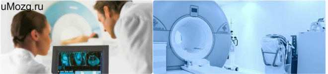
> CT and X-ray
Diagnosis of diseases using x-rays is still very relevant in modern medicine. It helps to study the structure internal organs, soft tissues and bones. Modern x-rays have taken on a variety of guises, some of which are almost unrecognizable. Classical radiography is used to diagnose bones, jaws and joints. As a rule, it is indispensable for detecting fractures, cracks. This is a linear scan that creates an image by "running" a beam of rays through the area being examined once.
To convert the image to numeric values, the image has been subdivided into a grid containing a number of pixels, which are used as a unit of measure to describe the geometric dimension of the image. Each pixel carries information about the color or gray level it represents.
Contraindications for CT
One way to observe the distribution of gray levels in an image is through a histogram. In the gray scale, black is represented by 0 and white by 255. Thus, each image provides a specific tone composition, depending on the attenuation coefficient of each soil particle.
Computed tomography is also a scan using x-rays. However, the images are created slice-by-slice in different planes. This allows you to look at the same organ from different angles, due to which the diagnostic possibilities increase significantly. At the same time, it is possible to differentiate soft tissues, the organs are not superimposed on each other, but are depicted separately. In the images, even structures with a difference in density of no more than 0.1% can be distinguished, which is impossible with conventional x-rays.
To validate the proposed methodology, a Phantom was used to physically characterize the template to test the pore separation threshold and solids. Within the largest circle, there are nine spheres not filled with solid material that made noise, and there is a lot of noise near the edge of the largest circle.
The image processing research used several methods to improve the image and obtain the information needed to characterize it. The analysis methods were based on mathematical morphology. Mathematical morphology is a tool used to extract image components that are useful in representing and describing the shape of an area.
When is X-ray used?
But despite all the benefits computed tomography, it cannot be completely displaced by the classical X-ray. This is due to many reasons. The fact is that the x-ray:
- More affordable - equipment is available in most clinics in the country, including state clinics.
- Cheaper - the cost for an x-ray check is from 300 to 1500 rubles, while for a tomography you will have to pay at least 3-4 thousand.
- Safer - the radiation dosage is 10-15 times lower (0.2-0.9 mSv) than with computed tomography (3-10 mSv). This allows you to do repeated scans without unnecessary fears for human health with an interval of several days.
X-rays best show bone tissue, so it is used to detect fractures, cracks, and other bone pathologies. However, this technique does not provide information about the structure of the bones, does not allow you to look inside the studied part of the body.
Of the ten tomographic images, an overlay was performed, and its resulting image is shown in Figure 7. This shows that simply overlaying images without accepting appropriate criteria is not enough to improve the final image quality and may even degrade it. However, this study focused on the edge of the image, a result in which the criteria used were effective.
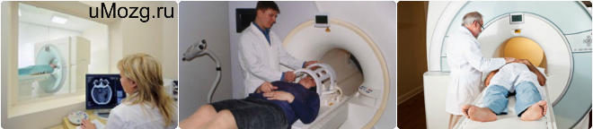
CT scans are performed in our center
The second stage of the analysis was to change the shape of the structuring element. A disk shape representing a round shape has been chosen. After determining the shape and size of the structuring element, the morphological operations of opening and closing were performed. Opening is a morphological operation that usually smoothes the contour of the image, breaks narrow bridges and eliminates thin protrusions. The close operation will fill or close the voids, removing many pixels of white noise.
When to Avoid CT
If the results of radiography do not provide sufficient information about the condition of the bones, soft tissues and do not allow a conclusion about the disease, then a CT scan is prescribed. As a rule, it happens, if necessary, to examine:
- Complex structures - skull, inner ear, teeth
- Abdominal organs - intestines, stomach, lungs, bladder
- Tumors and metastases - to accurately identify their localization and characterization
The main advantage of computed tomography is that it allows you to simultaneously assess the condition of bones, soft tissues, and blood vessels. The additional use of contrast greatly expands the possibilities of this technique. The accuracy of the obtained data reaches 97-98%. As a rule, CT provides a complete picture of the anatomical processes in the body, reveals the diagnosis and prescribes the appropriate treatment.
Computed X-ray tomography. Principles of obtaining computed tomograms. Features of the image of organs and tissues
The third stage of the analysis included the localization of the control point to separate the background image of the object of study. The method used is based on the calculation of the Euclidean distance given. Euclidean distance is a procedure that allows you to calculate the pixel distance of the nearest pixel background belonging to an object inside an image from the image containing the object.
Indications for computed tomography
In this study, three methodologies were adopted to determine the threshold values in the original tomographic image. The first methodology was to accept a 5% error over the range of gray levels present in the image, called the "5% Error Method".
What to choose?
Since the diagnostic capabilities of radiography are rather limited, it makes no sense to carry out this procedure instead of computed tomography. But the reverse replacement is possible - the tomogram will show all the same pathologies and changes as in a conventional x-ray. But it makes no sense to thoughtlessly conduct a CT scan instead of an X-ray. If the task comes down to assessing the consequences of an injury, diagnosing a fracture, dislocation or fracture of a bone, then there is no point in exposing your body to excess radiation exposure. All this will be perfectly visible with the help of a conventional x-ray.
The second methodology used a separatrix measurement called percentiles. This statistic is based on calculating the percentiles of the image histogram frequency distribution. The value of the first percentile calculated at 5% is accepted. This is a method that automatically selects the gray level limits that best separate or segment elements of interest in a particular class of image, with selection criteria that minimize within-group variance. When separating the value, which is considered porous or solid, the images were binarized.
As a rule, if it is necessary to examine the skeleton, an x-ray is first prescribed. There is almost no harm from it, but it is likely that the testimony of this diagnosis will be enough. If the data obtained is too small to make a diagnosis, then an additional computed tomography is prescribed, which will allow you to see hidden pathologies. In a word, the choice of CI or X-ray is incorrect. When making a decision, be guided by the opinion of a specialist who prescribes an appointment, taking into account the alleged diagnosis and the estimated radiation exposure.
Binarization was performed by sweeping the image, pixel by pixel, and applying Equation 2. Converting the image with gray levels to binary representation is important to calculate the percentage of pores and particulates present in the image. After determining the threshold value, the percentile method was applied on eleven images selected from the central part of the undisturbed soil sample in the three soil management systems assessed.
Threshold methods have shown excellent results. The percentile method was the one that presented results closer to the link. The 5% error method overestimated the solid image values, while the Otsu method underestimated the values. It is observed in the image that the method with 5% error generates a lot of noise in small circles. With regard to the Otsu method, the opposite result was obtained with a predominance of noise inside the largest circle.
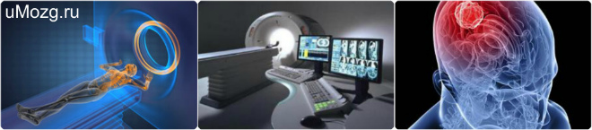
CT or X-ray computed tomography (CT) is one of the most accurate methods for diagnosing diseases. This method is characterized by the measurement of the X-ray attenuation coefficient when passing through different tissues and the possibility of layer-by-layer diagnostics of the structure inside the object.
The method has the ability of high contrast resolution, which allow differentiating tissues with different percentage density. It also allows you to give accurate information about the localization and nature of the pathology, its effect on the nearest structures.
The method that provided greater accuracy was the percentile method, which had a threshold value intermediate to the other methods. There are various studies on the problem of image segmentation in soils, and one conclusion that can be drawn from them is that a simple simple best segmentation method is not currently available, and the choice of an appropriate procedure depends largely on image quality, resolution, frequency the distribution of attenuation values and, finally, the focus of the study.
Indications for the CT procedure
The macropore and solids percentage values showed no difference between soil formation without tillage and conventional tillage. However, compared with the results obtained in the forest zone, a 10-fold increase is observed in the percentage of soil pores.
The CT image today shows a fully 3D image, which almost completely reduces the possibility of not detecting even minor pathologies.
Only a neurosurgeon or a neuropathologist is able to prescribe an CT scan of the brain, answer what it is and give the necessary recommendations. Diagnostics is performed in the following two groups:
Macroporosity is an important aspect of root development and hence plant development. Macroporosity values should be at least 10% of the total soil volume to allow gas exchange and root growth for most elevated crops. There was an average increase in soil macroporosity of about 22% in the area under forest in relation to the area without tillage and conventional tillage. These results confirm the rearrangement of soil particles contributing to tillage and conventional tillage.
How often can a CT scan be done?
Soil pore space, as assessed by tomographic and binarized images, was different for soil management with conventional tillage and no tillage in relation to forest area. Growing areas are dominated by micropores, which may be associated with greater extension, branching, and root activity. Grasses have tufts that penetrate the soil, giving the shape of a large matted, dense and voluminous.
- neuralgia of a different nature of development (transient, increasing or manifesting itself for the first time);
- With an increase in intracranial pressure;
- Convulsive and non-convulsive paroxysms (fainting, convulsive syndromes);
- Violation of cognitive functions (speech, memory, etc.);
- visual disturbances.
- According to nosological signs:
- Acute vascular pathology due to circulatory disorders in the brain, as well as the detection of ischemic and hemorrhagic stroke;
- craniocerebral;
- Primary tumor formations, as well as those formed as a result of metastasis, as well as after surgical intervention and treatment;
- Inflammatory diseases that have an acute and progressive course (abscess, encephalitis).
What is the difference between x-ray and tomography?
When observing the tomographic and binarized image in the forest zone, pores in the soil are visually more present, and the imaging technique is useful for detecting and confirming changes in soil macroporosity. The presence of holes of various sizes and shapes in the forest zone is observed. This change in soil structure affects water retention by influencing soil water dynamics.
It was found that soil management changed the soil porosity and that the methodology adopted could show that this difference was more pronounced in areas without tillage and conventional tillage compared to the area under forest.
What is CT of the brain, can be carried out using a special, so-called multispiral technology (MSCT). Which allows her to have advantages in the following cases:
- High scanning speed, which also allows you to get a complete image of the pathological area;
- The ability of MSCT to explore several areas at once;
- Significant improvement in contrast resolution;
- Advanced visualization lets you explore coronary arteries at almost any angle with obtaining their images, high definition;
- The possibility of conducting a study of patients who have built-in mechanical implants;
- Reduced radiation exposure from radiation pressure. The method is significantly safer than others that use X-rays.
A particularly important point should be noted that this method provides a reconstruction in a three-dimensional system. Therefore, this specialist, for example, reconstructive plastic surgery, works with an accurate picture of the area with which they have to work.
The method providing greater accuracy was the percentile method. Analysis of the tomographic image made it possible to distinguish soil porosity under various soil management conditions. The results are closer to reality when the scale used is related to soil macroporosity, however, other processes occur at the nanometer scale, which was not observed in this study.
Soil management changed soil porosity and this difference was more pronounced in areas without tillage and conventional tillage compared to the area under forest. The authors gratefully acknowledge the support of the Brazilian Agricultural Research Corporation, the coordination of the improvement of the quality of higher education staff, and the Research and Project Funding Authority for the financial support of these studies.
Carrying out diagnostics
The study of the pathological focus can be carried out with the help of the introduction of a contrast agent, as a rule, this is carried out to detect pathology in hard-to-reach areas, and without the introduction of contrast. Contrasting allows you to reproduce a more accurate image and accurately determine the desired area.
Precautions for the use of computed tomography
Compton tomography for agricultural measurements. Micromorphological features of soil geography. Threshold selection method from gray level histograms. The influence of root-induced biopores on the architecture of the pore space was studied using industrial X-ray computed tomography. Root Processes: Advances in Tomography and Imaging. Application of X-ray computed tomography for soil science: a review of the literature.
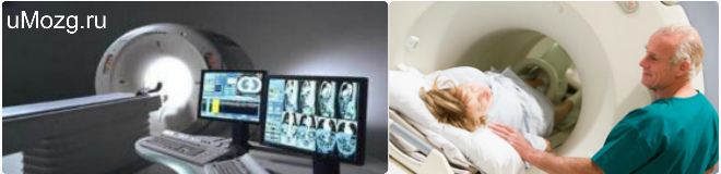
Invisible in the portal is an X-ray tube, the radiation of which is received by an invisible scanning unit in the gantry. Using a rotating thin X-ray beam, pieces are produced by the body, which are visually evaluated on a connected computer and combined to form an accurate image of the layer under study. This allows you to create cross-sectional images of the head and the entire body.
The doctor must identify all this study that the patient may have. Complete patient information and history should be the first decision to proceed with.
Any CT scan of the brain is not required, allowing immediate examination. The patient lies down on a moving transponder table, which then moves to the required point, depending on the area to be examined.
Is this method particularly suitable for organ systems?
We found individual images on a high resolution monitor. In some cases, it is necessary to administer an intravenous contrast agent containing iodine. The agent is generally well tolerated. When examining the digestive organs, depending on the question, an additional oral contrast agent is required.
The advantages over conventional "normal" X-ray images are better differentiation of soft tissues from each other, no bone overlap, and the ability to perform density measurements on individual structures. In addition, a cross-section of individual layers or a view of organs or structures at several levels is possible.
MSCT or MRI of the brain
To determine which of these methods is the most advantageous, it is necessary to determine their differences from each other. Based clinical manifestations the doctor determines the choice of diagnostic method:
- Systematic dizziness;
- Headache;
- Suspicion of the presence of a tumor;
- Symptoms of a stroke
- Traumatic brain injury;
- Developing deformity of the dentition.
How is the investigation going?
It can also be used with pacemakers. As a rule, it is better suited for examining the lungs. Other advantages are the short exam times and the usually short-term availability of the exam at emergency cases. Routine whole body examinations without and with contrast media Head and brain Sinuses Cervical soft tissue Lightweight respirators. In the preparation room, you will first be asked to remove jewelry, your watch, loose metal parts such as keys, removable dentures or hearing aids, your check card wallet, and the like.
To explore soft tissues, the state of blood circulation, in this case, the best way out is magnetic resonance imaging. However, CT is used in cases of diagnosis of bone tissue, sinuses. Experts do not undertake to argue which method is better, since each of them has its own contraindications and advantages.
Persons with metal implants and pacemakers are not allowed to conduct an MRI, as they can lead to equipment failure due to the magnetic field used. Computed tomography is contraindicated in pregnant women and women, as well as in persons who have recently undergone an x-ray.
The patient does not have the right to demand a referral to one or another method, since only a doctor is able to answer the patient's question, what is CT of the brain. To date, MRI is more expensive than CT, but soon they will be almost identical.
Rules for conducting CT (MSCT) of the brain
There is a certain set of rules on how to act before and during this diagnosis. Therefore, the following recommendations should be followed:
- The patient should lie comfortably with his back on the transponder table, while maintaining complete immobility. If this method is prescribed to a child or a patient with disorders in which he cannot remain still, a number of sedatives are introduced.
- The procedure does not take more than 15 minutes, except for the case with the introduction of a contrast agent;
- Metal objects are removed so that there is no possible distortion of the image;
- The possibility of carrying out the procedure for women in position exists only if this cannot be avoided;
- If the brain is being examined, then none is required;
- MSCT is also contraindicated in children due to the radiation received, but in some cases, diagnosis is still necessary;
When comparing CT with other similar methods (MRI, X-ray, and others), it is the method of resonance computed tomography that has the highest accuracy. One of the main disadvantages of CT is the increased risk of developing cancer during re-diagnosis, in the coming days after the first procedure.
Video