Antipyretics for children are prescribed by a pediatrician. But there are emergency situations for fever when the child needs to be given medicine immediately. Then the parents take responsibility and use antipyretic drugs. What is allowed to give to infants? How can you bring down the temperature in older children? What medicines are the safest?
Human circulation
Human blood circulation diagram
Human blood circulation- a closed vascular pathway that provides continuous blood flow, carrying oxygen and nutrition to cells, carrying away carbon dioxide and metabolic products. It consists of two successively connected circles (loops), starting from the ventricles of the heart and flowing into the atria:
- systemic circulation begins in the left ventricle and ends in the right atrium;
- pulmonary circulation begins in the right ventricle and ends in the left atrium.
Systemic (systemic) circulation
Structure
Functions
The main task of the small circle is gas exchange in the pulmonary alveoli and heat transfer.
“Additional” circulation circles
Systemic circulation video.
Both vena cavae bring blood to the right atrium, which also receives venous blood from the heart itself. This closes the circle of blood circulation. This blood path is divided into small and big circle circulation.

Pulmonary circulation video
Small circle of blood circulation(pulmonary) starts from the right ventricle of the heart with the pulmonary trunk, includes the branches of the pulmonary trunk to the capillary network of the lungs and the pulmonary veins flowing into the left atrium.
Systemic circulation(bodily) starts from the left ventricle of the heart with the aorta, includes all its branches, capillary network and veins of organs and tissues of the whole body and ends in the right atrium.
Consequently, blood circulation occurs through two interconnected circulation circles.
The regular movement of blood flow in circles was discovered in the 17th century. Since then, the study of the heart and blood vessels has undergone significant changes due to the acquisition of new data and numerous studies. Today, there are rarely people who do not know what the circulatory circles of the human body are. However, not everyone has detailed information.
In this review, we will try to briefly but succinctly describe the importance of blood circulation, consider the main features and functions of blood circulation in the fetus, and the reader will also receive information about what the circle of Willis is. The data presented will allow everyone to understand how the body works.
Additional questions that may arise as you read will be answered by competent portal specialists.
Consultations are carried out online and free of charge.
In 1628, a physician from England, William Harvey, made the discovery that blood moves along a circular path - the systemic circulation and the pulmonary circulation. The latter includes blood flow to the lung respiratory system, and the large one circulates throughout the body. In view of this, the scientist Harvey is a pioneer and made the discovery of blood circulation. Of course, Hippocrates, M. Malpighi, as well as other famous scientists made their contribution. Thanks to their work, the foundation was laid, which became the beginning of further discoveries in this area.
general information
The human circulatory system consists of: the heart (4 chambers) and two circulatory circles.
- The heart has two atria and two ventricles.
- The systemic circulation begins from the ventricle of the left chamber, and the blood is called arterial. From this point, blood flows through the arteries to each organ. As they travel through the body, the arteries transform into capillaries, which exchange gases. Next, the blood flow turns into venous. Then it enters the atrium of the right chamber and ends in the ventricle.
- The pulmonary circulation is formed in the ventricle of the right chamber and goes through the arteries to the lungs. There the blood exchanges, giving off gas and taking up oxygen, exits through the veins into the atrium of the left chamber, and ends in the ventricle.
Diagram No. 1 clearly shows how the blood circulation operates.
ATTENTION!
Many of our readers actively use a well-known method based on natural ingredients, discovered by Elena Malysheva, to treat HEART DISEASES. We recommend that you check it out.

It is also necessary to pay attention to the organs and clarify the basic concepts that are important in the functioning of the body.
The circulatory organs are as follows:
- atria;
- ventricles;
- aorta;
- capillaries, incl. pulmonary;
- veins: hollow, pulmonary, blood;
- arteries: pulmonary, coronary, blood;
- alveolus.
Circulatory system
In addition to the minor and major pathways of blood circulation, there is also a peripheral pathway.
Peripheral circulation is responsible for the continuous process of blood flow between the heart and blood vessels. The muscle of the organ, contracting and relaxing, moves blood throughout the body. Of course, the pumped volume, blood structure and other nuances are important. The circulatory system works due to the pressure and impulses created in the organ. The way the heart pulsates depends on the systolic state and its change to diastolic.
The vessels of the systemic circulation carry blood flow to organs and tissues.
- Arteries leaving the heart carry blood circulation. Arterioles perform a similar function.
- Veins, like venules, help return blood to the heart.
Arteries are tubes through which a large circle of blood flows. They have a fairly large diameter. Able to withstand high pressure due to thickness and ductility. They have three shells: inner, middle and outer. Thanks to their elasticity, they independently regulate depending on the physiology and anatomy of each organ, its needs and the temperature of the external environment.
The system of arteries can be imagined as a bush-like bundle, which becomes smaller the further from the heart. As a result, in the limbs they look like capillaries. Their diameter is no larger than a hair, and they are connected by arterioles and venules. Capillaries have thin walls and have one epithelial layer. This is where the exchange of nutrients takes place.
Therefore, the importance of each element should not be underestimated. Violation of the functions of one leads to diseases of the entire system. Therefore, to maintain the functionality of the body, you should lead a healthy lifestyle.
Heart third circle
As we found out, the pulmonary circulation and the large circulation are not all components of the cardiovascular system. There is also a third path along which blood flow occurs and it is called the cardiac circulation circle.

This circle originates from the aorta, or rather from the point where it divides into two coronary arteries. The blood penetrates through them through the layers of the organ, then through small veins it passes into the coronary sinus, which opens into the atrium of the chamber of the right section. And some of the veins are directed to the ventricle. The path of blood flow through the coronary arteries is called the coronary circulation. Together, these circles are a system that supplies blood and nutrients to the organs.
Coronary circulation has the following properties:
- increased blood circulation;
- supply occurs in the diastolic state of the ventricles;
- There are few arteries here, so dysfunction of one gives rise to myocardial diseases;
- excitability of the central nervous system increases blood flow.
Diagram No. 2 shows how the coronary circulation functions.

The circulatory system includes the little-known circle of Willis. Its anatomy is such that it is presented in the form of a system of vessels that are located at the base of the brain. Its importance is difficult to overestimate, because... its main function is to compensate for the blood that it transfers from other “pools”. The vascular system of the circle of Willis is closed.
Normal development of the Willis pathway occurs in only 55%. A common pathology is an aneurysm and underdevelopment of the arteries connecting it.
At the same time, underdevelopment does not affect the human condition in any way, provided that there are no violations in other pools. May be detected during MRI. An aneurysm of the arteries of the Willis circulation is performed as a surgical intervention in the form of its ligation. If the aneurysm has opened, the doctor prescribes conservative treatment methods.

The Willis vascular system is designed not only to supply blood flow to the brain, but also to compensate for thrombosis. In view of this, treatment of the Willis pathway is practically not carried out, because no health hazard.
Blood supply in the human fetus
The fetal circulation is the following system. Blood flow with a high content of carbon dioxide from the upper region enters the atrium of the right chamber through the vena cava. Through the hole, blood enters the ventricle and then into the pulmonary trunk. Unlike the human blood supply, the pulmonary circulation of the embryo does not go to the lungs, but to the duct of the arteries, and only then to the aorta.
Diagram No. 3 shows how blood flows in the fetus.

Features of fetal blood circulation:
- Blood moves due to the contractile function of the organ.
- Starting from the 11th week, breathing affects blood flow.
- Great importance is given to the placenta.
- The fetal pulmonary circulation does not function.
- Mixed blood flow enters the organs.
- Identical pressure in the arteries and aorta.
To summarize the article, it should be emphasized how many circles are involved in supplying blood to the entire body. Information about how each of them works allows the reader to independently understand the intricacies of the anatomy and functionality of the human body. Don't forget that you can ask a question at online mode and get an answer from competent specialists with medical education.
And a little about secrets...
- Do you often experience discomfort in the heart area (stabbing or squeezing pain, burning sensation)?
- You may suddenly feel weak and tired...
- Blood pressure keeps going up...
- There is nothing to say about shortness of breath after the slightest physical exertion...
- And you have been taking a bunch of medications for a long time, going on a diet and watching your weight...
But judging by the fact that you are reading these lines, victory is not on your side. That is why we recommend that you familiarize yourself with new technique of Olga Markovich, which has found an effective remedy for the treatment of HEART diseases, atherosclerosis, hypertension and cleansing of blood vessels.
Tests
27-01. In which chamber of the heart does the pulmonary circulation conventionally begin?
A) in the right ventricle
B) in the left atrium
B) in the left ventricle
D) in the right atrium
27-02. Which statement correctly describes the movement of blood through the pulmonary circulation?
A) begins in the right ventricle and ends in the right atrium
B) begins in the left ventricle and ends in the right atrium
B) begins in the right ventricle and ends in the left atrium
D) begins in the left ventricle and ends in the left atrium
27-03. Which chamber of the heart receives blood from the veins of the systemic circulation?
A) left atrium
B) left ventricle
B) right atrium
D) right ventricle
27-04. Which letter in the figure indicates the chamber of the heart in which the pulmonary circulation ends?
27-05. The picture shows the human heart and large blood vessels. What letter represents the inferior vena cava? 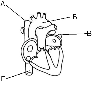
27-06. What numbers indicate the vessels through which venous blood flows?
A) 2.3
B) 3.4
B) 1.2
D) 1.4
27-07. Which statement correctly describes the movement of blood through the systemic circulation?
A) begins in the left ventricle and ends in the right atrium
B) begins in the right ventricle and ends in the left atrium
B) begins in the left ventricle and ends in the left atrium
D) begins in the right ventricle and ends in the right atrium
Circulation- this is the movement of blood through the vascular system, ensuring gas exchange between the body and the external environment, metabolism between organs and tissues and humoral regulation of various body functions.
Circulatory system includes the heart and - aorta, arteries, arterioles, capillaries, venules, veins etc. Blood moves through the vessels due to the contraction of the heart muscle.
Blood circulation occurs in a closed system consisting of small and large circles:
- The systemic circulation supplies all organs and tissues with blood and the nutrients it contains.
- The pulmonary, or pulmonary, circulation is designed to enrich the blood with oxygen.
Circulation circles were first described by the English scientist William Harvey in 1628 in his work “Anatomical Studies on the Movement of the Heart and Vessels.”
Small circle of blood circulation begins from the right ventricle, during the contraction of which venous blood enters the pulmonary trunk and, flowing through the lungs, gives off carbon dioxide and is saturated with oxygen. Oxygen-enriched blood from the lungs flows through the pulmonary veins into the left atrium, where the pulmonary circle ends.
Systemic circulation begins from the left ventricle, during the contraction of which blood enriched with oxygen is pumped into the aorta, arteries, arterioles and capillaries of all organs and tissues, and from there it flows through the venules and veins into the right atrium, where the great circle ends.
The largest vessel in the systemic circulation is the aorta, which emerges from the left ventricle of the heart. The aorta forms an arch from which arteries branch, carrying blood to the head () and to the upper extremities (vertebral arteries). The aorta runs down along the spine, where branches branch off from it, carrying blood to the abdominal organs, to the muscles of the trunk and lower extremities.
Arterial blood, rich in oxygen, passes throughout the body, delivering the nutrients and oxygen necessary for the cells of organs and tissues for their activities, and in the capillary system it turns into venous blood. Venous blood, saturated with carbon dioxide and products of cellular metabolism, returns to the heart and from it enters the lungs for gas exchange. The largest veins of the systemic circulation are the superior and inferior vena cava, which flow into the right atrium.
Rice. Diagram of the pulmonary and systemic circulation
You should pay attention to how the circulatory systems of the liver and kidneys are included in the systemic circulation. All blood from the capillaries and veins of the stomach, intestines, pancreas and spleen enters the portal vein and passes through the liver. In the liver, the portal vein branches into small veins and capillaries, which then reconnect into the common trunk of the hepatic vein, which flows into the inferior vena cava. All blood from the abdominal organs, before entering the systemic circulation, flows through two capillary networks: the capillaries of these organs and the capillaries of the liver. The portal system of the liver plays an important role. It ensures the neutralization of toxic substances that are formed in the large intestine during the breakdown of amino acids that are not absorbed in the small intestine and are absorbed by the colon mucosa into the blood. The liver, like all other organs, also receives arterial blood through the hepatic artery, which arises from the abdominal artery.
The kidneys also have two capillary networks: there is a capillary network in each Malpighian glomerulus, then these capillaries are connected to form an arterial vessel, which again breaks up into capillaries intertwining the convoluted tubules.

Rice. Circulation diagram
A feature of blood circulation in the liver and kidneys is the slowing down of blood flow, which is determined by the function of these organs.
Table 1. Differences in blood flow in the systemic and pulmonary circulation
|
Blood flow in the body |
Systemic circulation |
Small circle of blood circulation |
|
In which part of the heart does the circle begin? |
In the left ventricle |
In the right ventricle |
|
In which part of the heart does the circle end? |
In the right atrium |
In the left atrium |
|
Where does gas exchange occur? |
In the capillaries located in the thoracic and abdominal cavities, brain, upper and lower extremities |
In the capillaries located in the alveoli of the lungs |
|
What kind of blood moves through the arteries? |
Arterial |
Venous |
|
What kind of blood moves through the veins? |
Venous |
Arterial |
|
Time it takes for blood to circulate |
||
|
Circle function |
Supply of organs and tissues with oxygen and transport of carbon dioxide |
Saturation of blood with oxygen and removal of carbon dioxide from the body |
Blood circulation time - the time of a single passage of a blood particle through the major and minor circles of the vascular system. More details in the next section of the article.
Patterns of blood movement through vessels
Basic principles of hemodynamics
Hemodynamics is a branch of physiology that studies the patterns and mechanisms of blood movement through the vessels of the human body. When studying it, terminology is used and the laws of hydrodynamics are taken into account - the science of the movement of fluids.
The speed at which blood moves through the vessels depends on two factors:
- from the difference in blood pressure at the beginning and end of the vessel;
- from the resistance that the liquid encounters along its path.
The pressure difference promotes fluid movement: the larger it is, the more intense this movement. Resistance in vascular system, which reduces the speed of blood flow, depends on a number of factors:
- the length of the vessel and its radius (the longer the length and the smaller the radius, the greater the resistance);
- blood viscosity (it is 5 times greater than the viscosity of water);
- friction of blood particles against the walls of blood vessels and among themselves.
Hemodynamic parameters
The speed of blood flow in the vessels is carried out according to the laws of hemodynamics, common with the laws of hydrodynamics. The speed of blood flow is characterized by three indicators: volumetric speed of blood flow, linear speed of blood flow and blood circulation time.
Volumetric blood flow velocity - the amount of blood flowing through the cross section of all vessels of a given caliber per unit time.
Linear speed of blood flow - the speed of movement of an individual blood particle along a vessel per unit of time. In the center of the vessel, the linear velocity is maximum, and near the vessel wall it is minimum due to increased friction.
Blood circulation time - the time during which blood passes through the systemic and pulmonary circulation. Normally it is 17-25 s. It takes about 1/5 to pass through a small circle, and 4/5 of this time to pass through a large circle.
The driving force of blood flow in the vascular system of each circulatory system is the difference in blood pressure ( ΔР) in the initial section of the arterial bed (aorta for the great circle) and the final section of the venous bed (vena cava and right atrium). Blood pressure difference ( ΔР) at the beginning of the vessel ( P1) and at the end of it ( P2) is the driving force of blood flow through any vessel of the circulatory system. The force of the blood pressure gradient is used to overcome resistance to blood flow ( R) in the vascular system and in each individual vessel. The higher the blood pressure gradient in the blood circulation or in a separate vessel, the greater the volumetric blood flow in them.
The most important indicator of blood movement through the vessels is volumetric blood flow velocity, or volumetric blood flow(Q), which is understood as the volume of blood flowing through the total cross-section of the vascular bed or the cross-section of an individual vessel per unit time. Blood flow rate is expressed in liters per minute (l/min) or milliliters per minute (ml/min). To assess the volumetric blood flow through the aorta or the total cross-section of any other level of the vessels of the systemic circulation, the concept is used volumetric systemic blood flow. Since in a unit of time (minute) the entire volume of blood ejected by the left ventricle during this time flows through the aorta and other vessels of the systemic circulation, the concept of systemic volumetric blood flow is synonymous with the concept (IOC). The IOC of an adult at rest is 4-5 l/min.
Volumetric blood flow in an organ is also distinguished. In this case, we mean the total blood flow flowing per unit of time through all the afferent arterial or efferent venous vessels of the organ.
Thus, volumetric blood flow Q = (P1 - P2) / R.
This formula expresses the essence of the basic law of hemodynamics, which states that the amount of blood flowing through the total cross-section of the vascular system or an individual vessel per unit time is directly proportional to the difference in blood pressure at the beginning and end of the vascular system (or vessel) and inversely proportional to the resistance to flow blood.
The total (systemic) minute blood flow in the systemic circle is calculated taking into account the average hydrodynamic blood pressure at the beginning of the aorta P1, and at the mouth of the vena cava P2. Since in this section of the veins the blood pressure is close to 0 , then into the expression for calculating Q or MOC value is substituted R, equal to the average hydrodynamic arterial blood pressure at the beginning of the aorta: Q(IOC) = P/ R.
One of the consequences of the basic law of hemodynamics - the driving force of blood flow in the vascular system - is determined by the blood pressure created by the work of the heart. Confirmation of the decisive importance of blood pressure for blood flow is the pulsating nature of blood flow throughout cardiac cycle. During cardiac systole, when blood pressure reaches its maximum level, blood flow increases, and during diastole, when blood pressure is minimal, blood flow decreases.
As blood moves through the vessels from the aorta to the veins, blood pressure decreases and the rate of its decrease is proportional to the resistance to blood flow in the vessels. The pressure in arterioles and capillaries decreases especially quickly, since they have great resistance to blood flow, having a small radius, a large total length and numerous branches, creating an additional obstacle to blood flow.

The resistance to blood flow created in the entire vascular bed of the systemic circulation is called total peripheral resistance(OPS). Therefore, in the formula for calculating volumetric blood flow, the symbol R you can replace it with an analogue - OPS:
Q = P/OPS.
From this expression a number of important consequences are derived that are necessary for understanding the processes of blood circulation in the body, assessing the results of measuring blood pressure and its deviations. Factors influencing the resistance of a vessel to fluid flow are described by Poiseuille’s law, according to which
![]()
Where R- resistance; L- length of the vessel; η - blood viscosity; Π - number 3.14; r- radius of the vessel.
From the above expression it follows that since the numbers 8 And Π are permanent L changes little in an adult, then the value of peripheral resistance to blood flow is determined by the changing values of the radius of blood vessels r and blood viscosity η ).
It has already been mentioned that the radius of blood vessels muscular type can change quickly and have a significant impact on the amount of resistance to blood flow (hence their name - resistive vessels) and the amount of blood flow through organs and tissues. Since resistance depends on the value of the radius to the 4th power, even small fluctuations in the radius of the vessels greatly affect the values of resistance to blood flow and blood flow. So, for example, if the radius of a vessel decreases from 2 to 1 mm, then its resistance will increase by 16 times and, with a constant pressure gradient, the blood flow in this vessel will also decrease by 16 times. Reverse changes in resistance will be observed when the radius of the vessel increases by 2 times. With a constant average hemodynamic pressure, blood flow in one organ can increase, in another - decrease, depending on the contraction or relaxation of the smooth muscles of the afferent arterial vessels and veins of this organ.
Blood viscosity depends on the content of the number of red blood cells (hematocrit), protein, lipoproteins in the blood plasma, as well as on the aggregate state of the blood. Under normal conditions, blood viscosity does not change as quickly as the lumen of blood vessels. After blood loss, with erythropenia, hypoproteinemia, blood viscosity decreases. With significant erythrocytosis, leukemia, increased erythrocyte aggregation and hypercoagulation, blood viscosity can increase significantly, which entails an increase in resistance to blood flow, an increase in the load on the myocardium and may be accompanied by impaired blood flow in the vessels of the microvasculature.
In a steady-state circulatory regime, the volume of blood expelled by the left ventricle and flowing through the cross-section of the aorta is equal to the volume of blood flowing through the total cross-section of the vessels of any other section of the systemic circulation. This volume of blood returns to the right atrium and enters the right ventricle. From it, blood is expelled into the pulmonary circulation and then returns to the pulmonary circulation through the pulmonary veins. left heart. Since the IOC of the left and right ventricles are the same, and the systemic and pulmonary circulations are connected in series, the volumetric velocity of blood flow in the vascular system remains the same.
However, during changes in blood flow conditions, for example when moving from a horizontal to a vertical position, when gravity causes a temporary accumulation of blood in the veins of the lower torso and legs, the MOC of the left and right ventricles may become different for a short time. Soon, intracardiac and extracardiac mechanisms regulating the work of the heart equalize the volume of blood flow through the pulmonary and systemic circulation.
With a sharp decrease in venous return of blood to the heart, causing a decrease in stroke volume, blood pressure may decrease. If it is significantly reduced, blood flow to the brain may decrease. This explains the feeling of dizziness that can occur when a person suddenly moves from a horizontal to a vertical position.
Volume and linear speed of blood flow in vessels
The total blood volume in the vascular system is an important homeostatic indicator. Its average value is 6-7% for women, 7-8% of body weight for men and is in the range of 4-6 liters; 80-85% of the blood from this volume is in the vessels of the systemic circulation, about 10% - in the vessels of the pulmonary circulation and about 7% - in the cavities of the heart.
The most blood is contained in the veins (about 75%) - this indicates their role in depositing blood in both the systemic and pulmonary circulation.
The movement of blood in the vessels is characterized not only by volume, but also linear speed of blood flow. It is understood as the distance a particle of blood moves per unit of time.
There is a relationship between the volumetric and linear velocity of blood flow, described by the following expression:
V = Q/Pr 2
Where V- linear blood flow velocity, mm/s, cm/s; Q- volumetric blood flow velocity; P- number equal to 3.14; r- radius of the vessel. Value Pr 2 reflects the cross-sectional area of the vessel.

Rice. 1. Changes in blood pressure, linear velocity of blood flow and cross-sectional area in various parts of the vascular system

Rice. 2. Hydrodynamic characteristics of the vascular bed
From the expression of the dependence of the linear velocity on the volume in the vessels of the circulatory system, it is clear that the linear velocity of blood flow (Fig. 1) is proportional to the volumetric blood flow through the vessel(s) and inversely proportional to the cross-sectional area of this vessel(s). For example, in the aorta, which has the smallest cross-sectional area in the systemic circulation (3-4 cm2), linear speed of blood movement the largest and at rest is about 20-30 cm/s. With physical activity it can increase 4-5 times.
Towards the capillaries, the total transverse lumen of the vessels increases and, consequently, the linear speed of blood flow in the arteries and arterioles decreases. In capillary vessels, the total cross-sectional area of which is greater than in any other section of the vessels of the great circle (500-600 times larger than the cross-section of the aorta), the linear velocity of blood flow becomes minimal (less than 1 mm/s). Slow blood flow in capillaries creates the best conditions for metabolic processes between blood and tissues. In the veins, the linear velocity of blood flow increases due to a decrease in their total cross-sectional area as they approach the heart. At the mouth of the vena cava it is 10-20 cm/s, and with loads it increases to 50 cm/s.
The linear speed of plasma movement depends not only on the type of vessel, but also on their location in the blood flow. There is a laminar type of blood flow, in which the flow of blood can be divided into layers. In this case, the linear speed of movement of the layers of blood (mainly plasma) close or adjacent to the wall of the vessel is the lowest, and the layers in the center of the flow are the highest. Friction forces arise between the vascular endothelium and the parietal blood layers, creating shear stresses on the vascular endothelium. These tensions play a role in the endothelium’s production of vasoactive factors that regulate the lumen of blood vessels and the speed of blood flow.
Red blood cells in blood vessels (with the exception of capillaries) are located predominantly in the central part of the blood flow and move in it at a relatively high speed. Leukocytes, on the contrary, are located predominantly in the parietal layers of the blood flow and perform rolling movements at low speed. This allows them to bind to adhesion receptors in places of mechanical or inflammatory damage to the endothelium, adhere to the vessel wall and migrate into tissues to perform protective functions.
With a significant increase in the linear speed of blood movement in the narrowed part of the vessels, in the places where its branches depart from the vessel, the laminar nature of blood movement can be replaced by turbulent one. In this case, the layered movement of its particles in the blood flow may be disrupted; greater frictional forces and shear stresses may arise between the vessel wall and the blood than during laminar movement. Eddy blood flows develop, increasing the likelihood of damage to the endothelium and deposition of cholesterol and other substances into the intima of the vessel wall. This can lead to mechanical disruption of the structure of the vascular wall and initiation of the development of wall thrombi.
Time of complete blood circulation, i.e. the return of a blood particle to the left ventricle after its ejection and passage through the systemic and pulmonary circulation is 20-25 seconds per mow, or after approximately 27 systoles of the ventricles of the heart. Approximately a quarter of this time is spent moving blood through the vessels of the pulmonary circulation and three quarters through the vessels of the systemic circulation.
Encyclopedic YouTube
1 / 5
✪ Circulation circles. Big and small, their interaction.
✪ Circulatory circles, easy diagram
✪ Human blood circulation circles in 60 seconds
✪ Structure and work of the heart. Circulation circles
✪ Two circles of blood circulation
Subtitles
Systemic (systemic) circulation
Structure
Functions
The main task of the small circle is gas exchange in the pulmonary alveoli and heat transfer.
“Additional” circulation circles
Depending on the physiological state of the body, as well as practical expediency, additional circles of blood circulation are sometimes distinguished:
- placental
- cordial
Placental circulation
The mother's blood enters the plakura, where it gives oxygen and nutrients to the capillaries of the fetal umbilical vein, which runs along with two arteries in the umbilical cord. The umbilical vein gives off two branches: most of the blood flows through the ductus venosus directly into the inferior vena cava, mixing with unoxygenated blood from the lower part of the body. A smaller portion of the blood enters the left branch of the portal vein, passes through the liver and hepatic veins and then also enters the inferior vena cava.
After birth, the umbilical vein empties and turns into the round ligament of the liver (ligamentum teres hepatis). The ductus venosus also turns into a scar cord. In premature infants, the ductus venosus may function for some time (it usually becomes scarred after some time. If not, there is a risk of developing hepatic encephalopathy). In portal hypertension, the umbilical vein and the Arantian duct can recanalize and serve as bypass pathways (porto-caval shunts).
Mixed (arterial-venous) blood flows through the inferior vena cava, the oxygen saturation of which is about 60%; Venous blood flows through the superior vena cava. Almost all the blood from the right atrium flows through the foramen ovale into the left atrium and then into the left ventricle. From the left ventricle, blood is ejected into the systemic circulation.
A smaller portion of the blood flows from the right atrium into the right ventricle and pulmonary trunk. Since the lungs are in a collapsed state, the pressure in the pulmonary arteries is greater than in the aorta, and almost all the blood passes through the ductus arteriosus into the aorta. The ductus arteriosus flows into the aorta after the arteries of the head and upper extremities depart from it, which provides them with more enriched blood. A very small part of the blood enters the lungs, which subsequently enters the left atrium.
Part of the blood (about 60%) from the systemic circulation enters the placenta through the two umbilical arteries of the fetus; the rest goes to the organs of the lower body.
With a normally functioning placenta, the blood of the mother and fetus never mixes - this explains the possible difference in blood groups and Rh factor of the mother and fetus(es). However, determining the blood type and Rh factor of a newborn child from umbilical cord blood is often erroneous. During the birth process, the placenta experiences “overload”: pushing and the passage of the placenta through the birth canal contribute to pushing maternal blood into the umbilical cord (especially if the birth took place “unusually” or there was a pregnancy pathology). To accurately determine the blood type and Rh factor of a newborn, blood should be taken not from the umbilical cord, but from the child.
Blood supply to the heart or coronary circulation
It is part of a large circle of blood circulation, but due to the importance of the heart and its blood supply, you can sometimes find mention of this circle in the literature.
Arterial blood enters the heart through the right and left coronary arteries, originating from the aorta above its semilunar valves. The left coronary artery is divided into two or three, rarely four arteries, of which the most clinically significant are the anterior descending (LAD) and circumflex branches (OB). The anterior descending branch is a direct continuation of the left coronary artery and descends to the apex of the heart. The circumflex branch departs from the left coronary artery at its beginning at approximately a right angle, bends around the heart from front to back along the left edge of the heart, sometimes reaching the posterior wall of the interventricular groove. The arteries enter the muscle wall, branching to the capillaries. The outflow of venous blood occurs mainly into 3 veins of the heart: large, middle and small. Merging, they form the coronary sinus, which opens into the right atrium. The rest of the blood flows through the anterior cardiac veins and the Tebasian veins.
compensation for insufficient blood supply. Normally, the circle of Willis is closed. The formation of the circle of Willis involves the anterior communicating artery, the initial segment of the anterior cerebral artery (A-1), the supraclinoid part of the internal carotid artery, the posterior communicating artery, initial segment of the posterior cerebral artery(P-1).
In our body blood continuously moves through a closed system of blood vessels in a strictly defined direction. This continuous movement of blood is called blood circulation. Circulatory system a person is closed and has 2 circles of blood circulation: large and small. The main organ that ensures blood movement is the heart.
The circulatory system consists of hearts And vessels. There are three types of vessels: arteries, veins, capillaries.
Heart- a hollow muscular organ (weight about 300 grams) approximately the size of a fist, located in the chest cavity on the left. The heart is surrounded by a pericardial sac formed by connective tissue. Between the heart and the pericardial sac there is a fluid that reduces friction. Humans have a four-chambered heart. The transverse septum divides it into left and right halves, each of which is separated by valves, neither the atrium nor the ventricle. The walls of the atria are thinner than the walls of the ventricles. The walls of the left ventricle are thicker than the walls of the right, as it makes great job, pushing blood into the systemic circulation. At the border between the atria and ventricles there are leaflet valves that prevent the reverse flow of blood.
The heart is surrounded by the pericardium (pericardium). The left atrium is separated from the left ventricle by the bicuspid valve, and the right atrium from the right ventricle by the tricuspid valve.
Strong tendon threads are attached to the valve leaflets on the ventricular side. This design prevents blood from moving from the ventricles into the atrium during ventricular contraction. At the base of the pulmonary artery and aorta are semilunar valves that prevent blood from flowing from the arteries back into the ventricles.
The right atrium receives venous blood from the systemic circulation, and the left atrium receives arterial blood from the lungs. Since the left ventricle supplies blood to all organs of the systemic circulation, the left ventricle supplies arterial blood from the lungs. Since the left ventricle supplies blood to all organs of the systemic circulation, its walls are approximately three times thicker than the walls of the right ventricle. Cardiac muscle is a special type of striated muscle in which the muscle fibers grow together at their ends and form a complex network. This structure of the muscle increases its strength and speeds up the passage nerve impulse(the whole muscle reacts simultaneously). Cardiac muscle differs from skeletal muscles in its ability to contract rhythmically in response to impulses originating in the heart itself. This phenomenon is called automaticity.
arteries- vessels through which blood moves from the heart. Arteries are thick-walled vessels middle layer which are represented by elastic and smooth muscles, so the arteries are able to withstand significant blood pressure and not rupture, but only stretch.


The smooth muscles of the arteries perform not only a structural role, but its contractions contribute to the fastest flow of blood, since the power of the heart alone would not be enough for normal blood circulation. There are no valves inside the arteries; blood flows quickly.
Vienna- vessels that carry blood to the heart. The vein walls also have valves that prevent blood from flowing back.
Veins are thinner-walled than arteries, and the middle layer has fewer elastic fibers and muscle elements.
Blood through the veins does not flow entirely passively; the surrounding muscles perform pulsating movements and drive blood through the vessels to the heart. Capillaries are the smallest blood vessels, through them blood plasma exchanges nutrients with tissue fluid. The capillary wall consists of a single layer of flat cells. The membranes of these cells have multi-membered tiny holes that facilitate the passage of substances involved in metabolism through the capillary wall.
Blood movement occurs in two circles of blood circulation.
Systemic circulation- this is the path of blood from the left ventricle to the right atrium: left ventricle aorta thoracic aorta abdominal aorta arteries capillaries in organs (gas exchange in tissues) veins superior (inferior) vena cava right atrium
Small circle of blood circulation– path from the right ventricle to the left atrium: right ventricle pulmonary trunk artery right (left) pulmonary capillaries in the lungs gas exchange in the lungs pulmonary veins left atrium
In the pulmonary circulation, venous blood moves through the pulmonary arteries, and arterial blood moves through the pulmonary veins after gas exchange in the lungs.
In the human body, the movement of blood through the systemic and pulmonary circulation is provided so that the liquid tissue successfully copes with its responsibilities: transporting substances necessary for their development to the cells and carrying away decay products. Despite the fact that such concepts as “large and small circle” are rather arbitrary, since they are not completely closed systems (the first goes into the second and vice versa), each of them has its own task and purpose in the work of the cardiovascular system.
The human body contains from three to five liters of blood (women have less, men have more), which continuously moves through the vessels. It is a liquid tissue that contains a huge number of different substances: hormones, proteins, enzymes, amino acids, blood cells and other components (their number is in the billions). Such a high content of them in plasma is necessary for the development, growth and successful functioning of cells.
Blood transmits nutrients and oxygen to tissues through capillary walls. Then it takes carbon dioxide and decay products from the cells and carries them to the liver, kidneys, and lungs, which neutralize them and remove them outside. If for some reason the blood flow is stopped, the person will die within the first ten minutes: this time is enough for the brain cells deprived of nutrition to die, and the body to be poisoned by toxins.
The substance moves through the vessels, which is a vicious circle consisting of two loops, each of which originates in one of the ventricles of the heart and ends in the atrium. Each circle has veins and arteries, and the composition of the substance that is in them is one of the differences between the circulatory circles.
The arteries of the large loop contain tissue enriched with oxygen, while the veins contain tissue saturated with carbon dioxide. In the small loop, the opposite picture is observed: blood that needs purification is in the arteries, while fresh blood is in the veins.

The small and large circles perform two different tasks in the functioning of the cardiovascular system. In a large loop, human plasma flows through the vessels, transfers the necessary elements to the cells and takes away waste. In a small circle, the substance is cleared of carbon dioxide and saturated with oxygen. In this case, the plasma flows through the vessels only forward: the valves prevent the reverse movement of the liquid tissue. This system, consisting of two loops, allows different types blood do not mix with each other, which greatly facilitates the task of the lungs and heart.
How is blood purified?
The functioning of the cardiovascular system depends on the work of the heart: contracting rhythmically, it forces blood to move through the vessels. It consists of four hollow chambers located one after another according to the following scheme:
- right atrium;
- right ventricle;
- left atrium;
- left ventricle
Both ventricles are significantly larger than the atria. This is due to the fact that the atria simply collect and send the substance that enters them into the ventricles, and therefore do less work (the right one collects blood with carbon dioxide, the left one – saturated with oxygen).
According to the diagram, the right side of the heart muscle does not touch the left. The small circle originates inside the right ventricle. From here, the blood with carbon dioxide is sent to the pulmonary trunk, which subsequently diverges in two: one artery goes to the right, the second to the left lung. Here the vessels are divided into a huge number of capillaries, which lead to the pulmonary vesicles (alveoli).

Further, gas exchange occurs through the thin walls of the capillaries: red blood cells, which are responsible for transporting gas through the plasma, detach carbon dioxide molecules from themselves and combine with oxygen (blood is transformed into arterial blood). Then the substance leaves the lungs through four veins and ends up in the left atrium, where the pulmonary circulation ends.
It takes the blood four to five seconds to complete the small circle. If the body is at rest, this time is enough to provide it with the required amount of oxygen. During physical or emotional stress, pressure on a person’s cardiovascular system increases, which causes blood circulation to accelerate.
Features of blood flow in a large circle
Purified blood enters from the lungs into the left atrium, then goes into the cavity of the left ventricle (this is where the systemic circulation begins). This chamber has the thickest walls, due to which, when contracted, it is able to eject blood with a force sufficient for it to reach the farthest parts of the body in a few seconds.

During contraction, the ventricle releases liquid tissue into the aorta (this vessel is the largest in the body). Then the aorta diverges into smaller branches (arteries). Some of them go up to the brain, neck, upper limbs, some go down and serve the organs that are located below the heart.
In the systemic circulation, the purified substance moves through the arteries. Their distinctive feature is elastic but thick walls. Then the substance flows into smaller vessels - arterioles, and from them into capillaries, whose walls are so thin that gases and nutrients easily pass through them.
When the exchange ends, the blood, due to the added carbon dioxide and breakdown products, acquires a darker color, transforms into venous blood and is sent through the veins to the heart muscle. The walls of the veins are thinner than the arterial ones, but are characterized by a large lumen, so they hold much more blood: about 70% of the liquid tissue is in the veins.
If the movement of arterial blood is mainly influenced by the heart, then venous blood moves forward due to the contraction of skeletal muscles, which pushes it forward, as well as breathing. Since most of the plasma in the veins moves upward, to prevent it from flowing in the opposite direction, the vessels are equipped with valves to hold it back. At the same time, the blood that flows to the heart muscle from the brain moves through veins that do not have valves: this is necessary to avoid blood stagnation.
Approaching the heart muscle, the veins gradually converge with each other. Therefore, only two large vessels enter the right atrium: the superior and inferior vena cava. A large circle is completed in this chamber: from here the liquid tissue flows into the cavity of the right ventricle, then gets rid of carbon dioxide.
The average speed of blood flow in a large circle when a person is in a calm state is a little less than thirty seconds. At physical exercise, stress, and other factors that excite the body, blood flow can accelerate, since the cells’ need for oxygen and nutrients during this period increases significantly.
Any diseases of the cardiovascular system negatively affect blood circulation, blocking blood flow, destroying vascular walls, which leads to starvation and cell death. Therefore, you need to be very careful about your health. If you experience pain in the heart, tumors in the limbs, arrhythmia and other health problems, be sure to consult a doctor so that he can determine the cause of circulatory disorders, malfunctions of the cardiovascular system and prescribe a treatment regimen.
After all, it’s a shame for future doctors not to know the basics - the blood circulation. Without having this information and an understanding of how blood moves through the body, it is impossible to understand the mechanism of development of vascular and heart diseases, to explain pathological processes, which flow into the heart with one or another lesion. Without knowing the blood circulation it is impossible to work as a doctor. This information will not hurt the average person either, because knowledge about one’s own body is never superfluous.
1 Big trip
To better understand how the systemic circulation works, let’s imagine a little? Let’s imagine that all the vessels of the body are rivers, and the heart is a bay, into the bay of which all the river channels flow. Let's go on a journey: our ship begins a long voyage. From the left ventricle we swim to the aorta - the main vessel of the human body. This is where the great circle of blood circulation begins.
Blood saturated with oxygen flows in the aorta, because aortic blood is distributed throughout the human body. The aorta gives off branches, like a river, tributaries that supply blood to the brain and all organs. Arteries branch to arterioles, which in turn give off capillaries. Bright, arterial blood gives oxygen and nutrients to cells, and takes away metabolic products of cellular life.
The capillaries are organized into venules, which carry dark, cherry-colored blood, because it has given oxygen to the cells. Venules collect into larger veins. Our ship completes its journey along the two largest “rivers” - the superior and inferior vena cava - and ends up in the right atrium. The journey is over. A large circle can be schematically represented as follows: the beginning is the left ventricle and the aorta, the end is the vena cava and the right atrium.
2 Small trip

What is the pulmonary circulation? Let's go on our second journey! Our ship originates from the right ventricle, from which the pulmonary trunk arises. Remember that when completing the systemic circulation, we moored in the right atrium? From it, venous blood flows into the right ventricle, and then, with cardiac contraction, is pushed into a vessel that extends from it - the pulmonary trunk. This vessel goes to the lungs, where it bifurcates into pulmonary arteries and then into capillaries.
Capillaries envelop the bronchi and alveoli of the lungs, give off carbon dioxide and metabolic products and are enriched with life-giving oxygen. Capillaries organize into venules as they exit the lungs and then into larger pulmonary veins. We are accustomed to the fact that venous blood flows in the veins. Just not in the lungs! These veins are rich in arterial, bright scarlet, O2-rich blood. Through the pulmonary veins, our ship sails into the bay, where its journey ends - in the left atrium.
So, the beginning of the small circle is the right ventricle and the pulmonary trunk, the end is the pulmonary veins and the left atrium. A more detailed description is as follows: the pulmonary trunk is divided into two pulmonary arteries, which in turn branch into a network of capillaries, like a web, encircling the alveoli, where gas exchange occurs, then the capillaries collect into venules and pulmonary veins, which flow into the left upper cardiac chamber of the heart.
3 Historical facts

Having dealt with the sections of the blood circulation, it seems that there is nothing complicated in their structure. Everything is simple, logical, understandable. Blood leaves the heart, collects metabolic products and CO2 from the cells of the whole body, saturates them with oxygen, venous blood returns to the heart, which, passing through the natural “filters” of the body - the lungs, becomes arterial again. But it took many centuries to study and understand the movement of blood flow in the body. Galen mistakenly assumed that the arteries contained air rather than blood.
This position today can be explained by the fact that in those days they studied vessels only on corpses, and in a dead body the arteries are bloodless, and the veins, on the contrary, are full of blood. It was believed that blood was produced in the liver, and was consumed in the organs. Miguel Servet in the 16th century suggested that “the spirit of life originates in the left cardiac ventricle, this is facilitated by the lungs, where the mixing of air and blood coming from the right cardiac ventricle occurs,” thus, the scientist recognized and described for the first time the small circle.
But practically no attention was paid to Servetus' discovery. Harvey is considered the father of the circulatory system, who already in 1616 wrote in his writings that the blood “circles throughout the body.” For many years he studied the movement of blood, and in 1628 he published a work that became a classic, and crossed out all Galen’s ideas about blood circulation; in this work, blood circulation circles were outlined.

Harvey did not discover only capillaries, discovered later by the scientist Malpighi, who supplemented the knowledge about the “circles of life” with a connecting capillary link between arterioles and venules. The scientist was helped to open the capillaries by a microscope, which provided magnification up to 180 times. Harvey's discovery was met with criticism and challenge by the great minds of those times, many scientists did not agree with Harvey's discovery.
But even today, reading his works, you are surprised at how accurately and in detail for that time the scientist described the work of the heart and the movement of blood through the vessels: “The heart, while doing work, first moves, and then rests in all animals while they are still alive. At the moment of contraction, it squeezes blood out of itself, the heart empties at the moment of contraction.” The blood circulation was also described in detail, except that Harvey could not observe the capillaries, but he accurately described that blood collects from the organs and flows back to the heart?
But how does the transition from arteries to veins occur? This question haunted Harvey. Malpighi revealed this secret of the human body by discovering capillary blood circulation. It’s a shame that Harvey did not live several years to see this discovery, because the discovery of capillaries confirmed with 100% certainty the veracity of Harvey’s teachings. The great scientist did not have the opportunity to feel the full triumph of his discovery, but we remember him and his enormous contribution to the development of anatomy and knowledge about the nature of the human body.
4 From largest to smallest

I would like to dwell on the main elements of the circulatory circles, which are their framework through which the blood moves - the vessels. Arteries are vessels that carry blood from the heart. The aorta is the most important and important artery of the body, it is the largest - about 25 mm in diameter, it is through it that blood flows to other vessels extending from it and is delivered to organs, tissues, and cells.
Exception: the pulmonary arteries do not carry O2-rich blood, but CO2-rich blood to the lungs.
Veins are vessels that carry blood to the heart, their walls are easily stretchable, the diameter of the vena cava is about 30 mm, and the diameter of small veins is 4-5 mm. Their blood is dark, the color of ripe cherry, rich in metabolic products.
Exception: the pulmonary veins are the only veins in the body through which arterial blood flows.
Capillaries are the thinnest vessels, consisting of only one layer of cells. The single-layer structure allows gas exchange, exchange of useful and harmful products between cells and directly capillaries.
The diameter of these vessels is only 0.006 mm on average, and the length is no more than 1 mm. That's how small they are! However, if we sum up the length of all the capillaries together, we get a very significant figure - 100 thousand km... Our body inside is shrouded in them like a cobweb. And it’s not surprising - after all, every cell of the body needs oxygen and nutrients, and capillaries can provide the supply of these substances. All vessels, both the largest and smallest capillaries, form a closed system, or rather two systems - the above-mentioned circulatory circles.
5 Important features

Why are blood circulation circles needed? Their role cannot be overestimated. Just as life on Earth is impossible without water resources, human life is impossible without the circulatory system. The main role of the large circle is:
- Providing oxygen to every cell of the human body;
- The flow of nutrients from the digestive system into the blood;
- Filtration of waste products from the blood into the excretory organs.
The role of the small circle is no less important than those described above: removing CO2 from the body and metabolic products.
Knowledge about the structure own body are never superfluous, knowledge of how the circulatory departments function leads to a better understanding of the work of the body, and also forms an idea of the unity and integrity of organs and systems, the connecting link of which is undoubtedly the bloodstream, organized in circulatory circles.


