Antipyretics for children are prescribed by a pediatrician. But there are emergency situations for fever when the child needs to be given medicine immediately. Then the parents take responsibility and use antipyretic drugs. What is allowed to give to infants? How can you bring down the temperature in older children? What medicines are the safest?
A bruise is a kind of closed, in which superficial soft tissues are damaged ( , subcutaneous fat, small vessels, muscles, etc.) bodies without violating their anatomical integrity ( i.e. without wound formation). Shoulder bruises are quite often combined with other types of injuries - bones, joints, muscle inflammation ( ), joints ( ), violation of the integrity of ligaments, tendons, nerve damage, etc.
Bad posture is common cause pain under the shoulder blade or shoulder blade. Over time, rounding your shoulders over your desk or steering wheel, overloading your shoulder with a heavy wallet, or reusing your arm can lead to tension and discomfort anywhere in your neck, shoulder, and upper back. By gently stretching in the doorway, you can loosen the muscles that go below your shoulder blades, relieving pressure and soreness. If constant and frequent stretching does not bring relief, and the pain interferes with your ability to function, talk to your doctor or physical therapist.
Shoulder bruises most often occur when falling on the shoulder area, colliding with solid objects, as well as after direct blows to the shoulder, heavy bodies falling on the shoulder, etc. The appearance of a shoulder bruise is always accompanied by intense, swelling at the site of injury, impaired mobility in one or multiple joints. On the skin at the site of injury, redness first appears, and then bruising ( ), petechiae ( intradermal petechial hemorrhages). With more severe bruises on the shoulder, hematomas may occur ( cavities filled with blood), impaired skin sensitivity.
Warm up with 5 to 10 minutes of light physical activity such as walking or jogging. Once you break the sweat, prepare your shoulder and upper back muscles for stretching with a series of head, shoulder, and arm circles. Your goal is to increase blood flow to the area you want to stretch, which makes the muscles looser and more pliable.
Stand in a doorway with your fingers pointing up to your nose and your arms relaxed at your sides. Align your head with your back, straighten your back and pull your shoulders back and down a little. Bend your right elbow and place your forearm on the wall to the right of the door frame. Position your elbow just below shoulder height. Align your forearm so that it runs perpendicular to the floor.
Shoulder Anatomy
 Shoulder - the initial part of the upper limb of a person. It is located between the shoulder and elbow joints of the arm. Like all other open areas of the body, the shoulder is covered on the outside with skin and subcutaneous fat. Muscles are located deeper than them. All muscles of the shoulder are divided into anterior ( coracobrachial, brachial, biceps, muscles) and back ( articular muscle of the elbow, triceps, ulnar muscle) groups. These muscles allow you to manipulate the forearm and make various movements in it. In addition to these muscles at the top of the shoulder, the muscles of the shoulder girdle are connected to it ( supraspinatus, infraspinatus, small round, deltoid, etc.), which, in turn, help the hand to perform certain actions in the shoulder joint.
 Deeper than the muscles in the shoulder region is one single bone ( brachial bone), which is connected at the top ( by head humerus ) with the shoulder blade, forms the shoulder joint. From below, it contacts the bones of the forearm ( radial and ulnar), forming elbow joint.
Step forward through the doorway into a lunge position with your left foot in front of your right. Keep your bent left knee directly over your left heel and point your toes forward. Keep your left hand at your waist on your left side. Hinge forward from your waist, pressing your chest straight forward over your left thigh. Let your knees bend slightly. When you feel a stretch point in your right shoulder area, hold it for at least 20 seconds. Repeat the exercise on the other side.
If you experience stiffness under your shoulder blades at work, take frequent breaks to stretch the area. Always move into the stretch slowly and deliberately while maintaining control of your movement. Breathe evenly throughout the stretch to help relax the muscles in your neck, shoulders, and upper back. Make adjustments as needed to improve the way you carry yourself - and reduce stress on your upper body. In addition to stretching, pay attention to your posture throughout the day. . A certified fitness instructor with decades of dance training, she has taught senior adults, teens and children.
The shoulder blade belongs to the bones of the shoulder girdle. Together with the clavicle, it connects the bones of the upper limb with the bones of the body.
Each joint ( elbow or shoulder) is covered on top with a connective tissue sheath called the articular capsule. This capsule limits the joint from other tissues, and also stabilizes the articular surfaces of the bones that form it. The articular surfaces of each of the bones are covered with hyaline cartilage, which provides optimal sliding between them and dampens movement in the joint.
She has written teaching and learning materials for several non-profit organizations, and her work has appeared in many major online publications. Fisk holds a Bachelor of Arts in Public and International Affairs from Princeton University. Serving tennis ball after tennis ball, sitting at a computer for long hours, or carrying a heavy backpack can cause the rhomboids, the shoulder girdle muscles located on the upper back, to become tight and painful.
Shaped like a diamond, the rhomboids connect your spine to your shoulder blades and help move your arms and shoulders, especially when raising your arm above your head. Excess muscle can cause pain between the shoulder blades; stretching can help release the rhomboids and relieve discomfort in the upper back.
In addition to the joint capsule, each joint is strengthened by a ligamentous apparatus. Joint ligaments are made up of strong connective tissue. They can be located both outside and inside the articular cavity of the joints. To bundles shoulder joint include three glenohumeral ligaments and a coracobrachial ligament. The elbow joint is firmly fixed by the ulnar and radial collateral ligaments, as well as the square and annular ligaments of the radius.
The main causes of pain in the shoulder with a bruise
Warm up with at least 10 minutes of light cardiovascular activity such as jogging, jump rope, or cycling. Perform one set of each of the push-ups and rows to activate your back muscles. Perform a seated stretch to release the rhomboids. Sit high in a chair with your feet flat on the floor and your knees tucked over your ankles; The height of the chair should allow the knees to bend at an angle of 90 degrees. Bring your inner knees and inner ankles together so that they touch each other. Cross your arms in front of your body and lean forward to rest your right elbow on your left knee and your left elbow on your right knee; keep your back straight.
Near ( above) with the shoulder joint is the acromioclavicular joint. This joint is the junction between the acromion ( bone process) scapula and the humeral end of the clavicle. This joint is strengthened by the coracoclavicular and acromioclavicular ligaments. The acromioclavicular joint is an inactive joint.
What to do for a shoulder injury?
Keep your elbows in place as you separate your knees; stop moving your knees when you feel a stretch between your shoulder blades. Hold the stretch for 15 seconds and release. Use a bar or railing to stretch your rhomboids. Stand against a stand that is securely attached to the wall. Grasp the bar just below your chest up with both hands, using an overhand grip and touching with your fingers. Place your feet 1 to 2 inches in front of the bar, thumbs touch. Keep your arms and legs in place as you push your hips back, which will pull your shoulders forward.
Oxygenated arterial blood enters the upper limbs through the axillary arteries. These arteries serve as a direct continuation of the subclavian arteries arising from the aorta ( right subclavian artery arises from the aortic arch through the brachiocephalic trunk). The aorta is the main main arterial vessel into which blood enters after passing.
Hold the stretch for 30 seconds. Stretch your arms forward into child's pose to release the rhomboids. Kneel on the floor or yoga mat with your thumbs touching with your toes and your knees separated by your hips. Sit on your heels and tilt your torso forward, resting it on your hips or, if possible, on the floor between your knees; place your forehead on the floor. Stretch your arms above your head and place your palms on the floor; you should feel a stretch in your upper back and neck. Press down on your palms to deepen the stretch.
Hold the child's pose for one to three minutes. Release your rhomboids during regular post-workout stretching sessions, as well as any time they have been stressed, such as after a tennis match or after using the computer. Check with your doctor about any rhomboid or back pain and before treating pain or injury on your own. Shoulder suture A bruised or bruised shoulder is a condition where the shoulder bone or shoulder joint is damaged or damaged, leading to the development of a purplish-bluish discoloration under the skin of the shoulder joint.
Both axillary arteries ( right and left) turn into brachial arteries that supply most of the shoulder. Thus, the main main vessels that bring blood to the tissues of the shoulder of each of the arms are the axillary and brachial arteries. They both pass along the medial ( inner side) of the hand surface. The axillary artery is localized in the axillary region, and the brachial artery is localized near the medial ( inner side) edges of the humerus.
Shoulder stiffness is a common complaint among athletes and people who participate in heavy physical activity such as lifting weights or lifting heavy boxes, etc. subcutaneous sinuses of the shoulder usually do not require treatment and heal on their own. It can take up to four to six weeks for the sinuses of the shoulder joint, which are periosteal in nature, to heal.
Types of sinus joint
Subcutaneous shoulder bruises are the least painful and heal quickly without any treatment. Shoulder bruises that are subcutaneous are superficial bruises and are also known as bruises. They develop when broken. blood vessels, which are present under the skin of the shoulder, resulting in a collection of blood under the surface of the skin. The sinus of the shoulder joint is purple or bluish-brown initially, and then it may turn green or yellow as it heals.
Venous blood from the shoulder area is removed through superficial ( lateral and medial subcutaneous) and deep veins. Superficial veins are located directly under the skin and subcutaneous fat. Deep veins pass under the muscles and adjoin the brachial artery throughout the shoulder. Superficial and deep veins in the upper region of the shoulder unite with each other, creating the so-called axillary vein, which further flows into the subclavian vein.
Shoulder bruises of this type usually heal on their own without requiring any treatment. Direct pressure on the affected shoulder joint causes pain and should be avoided. Intramuscular bruises on the shoulder or bruises on the muscles of the shoulder can be painful and take longer to heal than subcutaneous bruises. Intramuscular shoulder bruises occur when a blood vessel breaks and blood pools in the shoulder muscle under the skin. Intramuscular shoulder contusions result from a sharp jerk, blunt force injury, or torn muscle.
The color of these types of bruises is usually blue or bright purple. Intramuscular shoulder contusions are also larger than subcutaneous shoulder contusions. In some cases, hematoma formation occurs near or above the shoulder joint injury. If the patient has severe pain with bruising of the shoulder joint, attention should be paid to rule out a more serious injury to the shoulder joint.
All nerves that innervate the shoulder region ( and also shoulder girdle), depart from the brachial plexus. This plexus is formed by connecting several ( fifth, sixth, seventh, eighth, etc.) cervical spinal nerves. The branches of the brachial plexus are anatomically divided into two parts. That part of it, which is above the collarbone, is called supraclavicular. At this level, nerves emerge from the brachial plexus ( suprascapular, subclavian, etc.), innervating the muscles of the shoulder girdle, neck, chest and back.
Bone or periosteal bruising is usually the most painful, and the healing time required for this type of shoulder sinus is also very long. This is the outer layer of the humerus. If the cortex is severely damaged, then a fracture of the humerus can also occur, as well as bruising. Symptoms of periosteal bruising of the shoulder include sharp pain, intense swelling and extensive discoloration. The discoloration and swelling will gradually disappear over several weeks. Despite the reduction in pain, it can persist for more than two to three months.
Having given its supraclavicular branches above the clavicle, the brachial plexus then follows under it and passes from its inner side, heading towards the shoulder. Under the clavicle, subclavian branches depart from the brachial plexus, innervating various tissues ( muscles, skin, joints, etc.) of the upper limb, as well as certain muscles of the chest, shoulder girdle, back and chest. The shoulder is innervated by the lateral ( median nerve root, musculocutaneous nerve), rear ( radial and axillary nerves), medial ( medial cutaneous nerve of the shoulder, ulnar nerve, medial root of the median nerve) bundles of nerves branching from the brachial plexus.
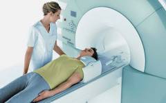
The pain, discoloration, and swelling that is present with periosteal bruising is usually severe enough that a fracture is suspected and needs medical attention. Some of the tests done for diagnosis include CT scan, magnetic resonance imaging. Plain x-rays are not helpful in detecting periosteal bruising; however, it is useful in detecting a fracture. If there is no fracture, then the diagnosis of hemoptysis is made by excluding a fracture, according to which an appropriate course of treatment is planned.
What structures can be damaged in a shoulder injury?
 With simple bruises, as a rule, more superficial layers are damaged - skin, subcutaneous fat, less often - muscles, their tendons or ligaments. With serious bruises, damage to deeper anatomical structures - joints, bones, nerves, blood vessels - often occurs.
 With a shoulder injury, the following anatomical structures can be damaged:
Causes of the sinus joint
Some of the common causes of shoulder bruises are trauma or trauma to the shoulder joint, a torn shoulder muscle, a sharp jerk or pull to the shoulder. Falling and landing on the shoulder joint can cause fracture and bruising. Heavy lifting can also lead to bruising of the shoulder joint.
Diagnosis of bruises on the shoulders
A bruised shoulder appears as a discoloration that is bluish-purple in color and may subsequently turn green-yellow during the healing phase. A patient with a shoulder bruise experiences pain and limited range of motion of the shoulder joint. If the injury that caused the bruising of the shoulder joint is severe, the patient may also experience pinched nerves, tingling fingers and cold hands. Immediate medical attention should be sought if the number of bruises is very large or if they swell rapidly.
- There is pain and swelling on the shoulder joint.
- There may be weakness in the muscles around the shoulder joint.
- There may also be a hematoma at the site of injury.
- Skin and subcutaneous tissue. The skin and subcutaneous tissue are the main structures that are damaged by bruises.
- Muscles. Muscles are often damaged by moderate to severe bruising. They can also be involved in various complications of shoulder contusion ( fractures, dislocations, damage to nerves, blood vessels, etc.).
- Muscular tendons. Muscle tendons are often damaged by contusions, complicated by fractures of the humerus or shoulder dislocations.
- Bones. Bones are usually damaged by contusions associated with their fractures or dislocations of the joints. With open fractures, suppuration in the bones may occur ( ) due to infection of the wound site with pathogens.
- joint structures. When a shoulder is bruised, a dislocation of the shoulder, an intra-articular fracture of the bones, or inflammation of the joint can sometimes occur. It is in these cases that damage to the articular structures occurs ( joint capsule, cartilage, bags, ligaments).
- Nerves and vessels. Nerves and blood vessels are often damaged by bruises due to their direct mechanical trauma ( impact, break, etc.) or because they are injured indirectly ( when mixing bone fragments with bone fractures, dislocation of the joint, etc.).
The main causes of pain in the shoulder with a bruise
 A bruise is one of the types of traumatic injury in which soft tissue damage occurs, which develops under the influence of an external force. Shoulder bruises are usually observed after direct violence, that is, after blows to this area, falls on it, collision of the shoulder with solid bodies, falling of something heavy on this zone. The severity of a bruise always depends on the mass, type, speed of the traumatic agent, as well as on the area of damage to the shoulder tissues.
 With a slight and superficial bruise of the shoulder, damage to the skin, small vessels and subcutaneous fat is noted, nerve endings and, to a lesser extent, muscles, ligaments. Quite often, small vessels at the site of injury can collapse. This results in minor bleeding in the skin that looks like petechiae ( petechial hemorrhages up to 1 - 2 mm in diameter). Petechiae in their size and predominant localization in the tissues differ from another type of hemorrhage - ecchymosis.
Sinus joint treatment
It is important to start treatment for shoulder bruises that are intramuscular or periosteal in nature. Timely treatment helps speed up the healing process, prevent complications and relieve pain. Shoulder bruising can be painful; however, it does not always require medical attention unless it is very severe. The following conservative methods can be done at home to relieve pain and discomfort from the sinus of the shoulder joint.
Pressure: Applying gentle pressure immediately after an injury helps slow or stop bleeding, thereby preventing bruising or bruising. Rest: Rest and immobilization of the shoulder joint is important to heal the sinuses of the shoulder joint. To achieve this goal, you can use the shoulder canine, i.e. provide rest and help prevent further injury. A splint can also be used for immobilization, but make sure it is kept in place at all times.
Ecchymosis ( bruising or bruising) are significant subcutaneous hemorrhages. They develop in the area of subcutaneous fat. With them, it is saturated with blood, while the accumulation of blood as such does not occur in it. Often with ecchymosis, skin and / or muscles can be impregnated along with subcutaneous fat. In some cases, with severe bruising of the shoulder, blood can accumulate in the subcutaneous fat, forming a hematoma ( cavity inside tissue filled with blood).
Bruises after a shoulder injury are very well recognized on the 2nd - 3rd day in the form of blue ( blue purple) spots ( in some cases, they may appear within minutes or hours of injury). As blood elements decay in the tissues, these spots acquire a green and then a yellow tint. A change in color from blue to green or yellow begins to occur from 4 to 10 days after the injury. Until then, the site of injury usually remains cyanotic. With a slight and superficial bruise of the shoulder, the bruises resolve after 12 to 16 days.
The characteristic signs of a shoulder injury are also considered the appearance of swelling and pain at the site of injury. Pain in a shoulder injury is usually caused either by direct damage to the nerve endings located in the tissues of the shoulder, or by their compression due to increasing swelling of these tissues ( nerves can be compressed and hematoma). Edema ( swelling) of shoulder tissues is caused by their inflammation. Inflammation is a pathological response of the body to damage or destruction of its tissues. It is accompanied by a local expansion of small vessels, an increase in their permeability, an increase in blood flow to the affected tissues. All this is the reason why they develop.
With severe bruises of the shoulder region, deeper structures can also be damaged ( bones, joints, ligaments, muscles). Therefore, so often shoulder bruises are associated with other types of shoulder injuries. The presence of an additional type of injury contributes to the occurrence of even more severe pain and even more pronounced swelling of the shoulder.
Shoulder bruises can be combined with the following types of injuries:
- fracture of the humerus, scapula, collarbone;
- dislocation of the shoulder, collarbone;
- muscle inflammation;
- inflammation of the shoulder or elbow joint;
- tendon injury;
- damage peripheral nerves;
- open shoulder injury.
Fracture of the humerus, scapula, clavicle
Fracture of the humerus, scapula, collarbone is a common occurrence with bruises in the shoulder region. With fractures, partial or complete destruction of the anatomical structure of the bones occurs. Such fractures usually occur when falling on the elbow joint, shoulder, or with a direct blow to the shoulder region. Fracture of these bones is characterized by the appearance severe pain and edema in the area of damage, as well as the occurrence of pathological mobility ( the presence of mobility in the place where it is not normally detected), crepitations ( crunching of bone fragments), joint dysfunction ( elbow, shoulder), shortening of the shoulder. Often, due to severe pain, the patient cannot move the injured arm, so he is forced to keep it healthy.The pain that develops with a fracture is usually much stronger than the pain caused by a bruise. However, unlike a fracture, with a bruise, the function of the joint is rarely severely impaired, and there should be pathological mobility, shortening of the shoulder and crepitus at all.
The problem is that the patient these symptoms ( crepitus, abnormal mobility) cannot always identify on its own, due to the existence various types closed fractures of these bones. Therefore, if there is severe pain in the shoulder or elbow joint that occurs immediately after a shoulder injury, all patients should definitely consult a doctor.
Shoulder dislocation, collarbone
A dislocation is a pathological situation in which the bones in the joints are separated from each other. With bruises of the shoulder, it is quite common to find dislocations of the shoulder or collarbone. They are observed when patients fall on the elbow, shoulder, direct blows to the area of the shoulder or acromioclavicular joints. With these dislocations, patients complain of sharp local pain in the area of the damaged joint, its swelling, and shoulder deformity. If there is a dislocation of the shoulder joint, the patient also makes additional complaints about the inability to carry out any movements in this joint. With dislocations of the shoulder or collarbone, the affected arm is supported by the injured with the help of a healthy one. Pain with such dislocations, they are caused by damage to the articular capsule, cartilage, ligament rupture, damage to the periosteum, compression of adjacent nerves and vessels.muscle inflammation
When the shoulder is bruised, not only the skin and subcutaneous fat are injured and inflamed, but sometimes the skeletal muscles as well. Inflammation of skeletal muscles ( myositis) develops for the same reason as the bruise itself ( due to mechanical injury), so myositis often accompanies bruises. Inflammation of the muscles during bruises, as a rule, is limited to the site of damage, it does not spread to neighboring tissues. With myositis in the damaged area of the shoulder, muscle pain and swelling in the muscles occur, mobility in the joints is limited ( due to pain in the muscles that regulate movement in it). Pain in myositis is aggravated by the slightest involvement of the affected muscles in movements, as well as by palpation of the site of injury.Sprain
Ligament sprain is a situation in which part of their connective tissue fibers is torn. Since ligaments are a component of the articular apparatus, sprains are usually observed in the area of \u200b\u200bthe joints ( shoulder or elbow) shoulder area. A sprain often accompanies bone fractures and dislocations that can occur with shoulder bruises. In the presence of only shoulder bruises, sprains are less common.Distinguish simple stretching ( that is, a sprain that developed on its own, without fracture or dislocation) from a shoulder injury that occurred after injuries of the shoulder area is practically impossible, since they clinical symptoms very similar. With a sprain, as well as with a bruise, pain, bruising, swelling at the site of injury, and limited mobility of the affected joint can occur.
Inflammation of the shoulder or elbow joint
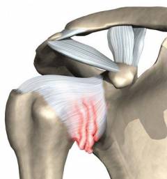
Shoulder injuries can damage joints ( elbow or shoulder). If their tissue is damaged ( articular cartilage, periosteum, articular capsule, etc.) become inflamed, due to which the joint increases in size, pathological fluid accumulates in it. With severe damage to the joints, small vessels can rupture in them, as a result of which blood will begin to accumulate in their joint cavity. This condition is called hemarthrosis. The appearance of hemarthrosis and the accumulation of pathological fluid in the joint cavity, as well as inflammation in its tissues, lead to a significant limitation of articular movements in the affected joints and the occurrence of intense pain.Tendon injury
Shoulder bruises, which are complicated by its dislocations, are often combined with ruptures of the muscle tendons. The most common type of injury that occurs in this case is a torn tendon of the rotator cuff. This cuff is a collection of several muscles ( infraspinatus, supraspinatus, subscapular, small round) shoulder girdle of the upper limb and their tendon bundles attached to the head of the humerus. The muscles that make up the rotator cuff are necessary to strengthen and stabilize the position of the humeral head in the shoulder joint. They are involved in abduction, adduction and rotational articular movements in the shoulder joint.When the tendons of these muscles are torn, there is a violation of movements in the shoulder joint. It causes pain and discomfort. Pain syndrome ( characteristic of rotator cuff tendon rupture) is often disguised as intense pain that develops as a result of dislocation and bruising. Therefore, in the early stages, such a gap is often not detected. It is usually diagnosed after the dislocation has been treated. As a rule, in patients with shoulder dislocation, pain and discomfort in the shoulder region does not completely disappear. This forces them to seek help again from their doctor, who, after conducting additional research, can identify a rupture of the tendons of the rotator cuff of the shoulder.
Peripheral nerve damage
For uncomplicated shoulder injuries without fractures, dislocations, etc.) may cause minor damage to the peripheral nerves of the hand. Most often, such injuries are manifested by pain, short-term numbness of the limb, tingling sensation, impaired motor ( motor) functions of certain muscles, redness or blanching of the skin on the shoulder. If a shoulder injury occurs simultaneously with a fracture or dislocation, then nerve damage can be very significant ( muscle paralysis, persistent disorders of skin sensitivity, dysregulation of vascular tone, etc.).With serious injuries of the shoulder, the axillary, subscapular, ulnar, musculocutaneous and radial nerves can most often be damaged. The first two nerves, for example, are often damaged by bruises, accompanied by dislocations of the shoulder or fractures of the head of the humerus. Their damage is characterized by loss of skin sensitivity on the upper lateral ( outer side) surface of the shoulder and paresis ( decrease in muscle strength) supraspinatus, infraspinatus ( subscapular nerve), small round, deltoid muscles ( axillary), as a result of which supination is disturbed ( outward rotation) and shoulder abduction.
Defeat radial nerve may be accompanied by a violation of skin sensitivity on the back of the shoulder, forearm and radial ( ray) hand surface, as well as paresis ( decrease in muscle strength) triceps brachii and extensor muscles of the hand. Because of such paresis, the hand will not be able to unbend in the wrist, and the forearm - in the elbow joint. Damage to this nerve usually occurs with bruises of the shoulder, complicated by closed fractures of the humerus in the area of its diaphysis ( middle part of the bone), lower metaphysis or epiphysis ( lower part of the bone).
When the musculocutaneous nerve is injured, paralysis occurs ( ) biceps, brachial and coracobrachial muscles, the sensitivity of the skin on the lateral ( outer side) side of the forearm. Due to such an injury, muscle weakness develops when the forearm is rotated outward and flexed at the elbow joint.
With fractures of the humerus in the region of its condyle, damage to the ulnar nerve often occurs. It is accompanied by a violation of the sensitivity of the skin on the elbow ( inner side) side of the forearm, hand and fingers ( in zone III, IV, V fingers). The fingers of the hand lose the ability to carry out various movements - adduction, flexion, extension, extension, etc.
Open shoulder injury
Shoulder contusion is often combined with bruised wounds. These wounds appear as a result of a blow to the shoulder region with a blunt object ( bottle, stick, metal rod, etc.). The edges of bruised wounds are always blurred, bleeding from them is very weak or absent at all ( since with them, basically, small superficial vessels are destroyed, which are immediately thrombosed). Pain and swelling occur at the site of injury. If the injury occurred in the area of the joints ( elbow, shoulder), then their function is partially violated.With more serious types open shoulder injury chopped, crushed, gunshot) there is unbearable pain, swelling and deformity of the shoulder, severe bleeding from the wound, the integrity of the superficial ( skin, subcutaneous fat) and deep tissues ( muscles, ligaments, tendons, bones, nerves, blood vessels), and with it the various functions of the hand.
What to do if you hit your shoulder hard?
With such bruises, it is recommended to drink anti-inflammatory drugs ( , dexalgin, etc.). There is no need to call an ambulance in such cases. However, it is still worth going to the emergency room for a consultation with a traumatologist so that he checks the safety of deeper anatomical formations ( ligaments, bones, joints).
Exactly the same first aid is provided if the shoulder injury occurred in the area of the shoulder or elbow joints of the arm. In these cases, a bandage pressure bandage may not be applied to the affected joint. Also, to reduce pain, it is recommended to move and feel the bruised place on the arm less. Appeal to the trauma department with a bruised joint must be carried out. Since, in most cases, with such injuries, pathological fluid accumulates in the cavity of damaged joints, which must be removed by articular puncture.
If, after a strong blow to the shoulder, a complicated shoulder injury appears, then in this case it is necessary to act on the basis of the situation. When a shoulder bruise is combined with a superficial wound, it must be treated with some kind of antiseptic ( alcohol, green) and apply a sterile bandage on top. After that, ice should be applied to the site of the bruise and an anesthetic should be used. Then you need to go to the emergency room.
- with fracture ( open and closed) or dislocation of the bones of the hand;
- with violation of the sensitivity of the skin of the hand ( shoulders, forearms, hands);
- with paresis ( weakening of muscle strength) or paralysis ( loss of a muscle's ability to contract) arm muscles;
- with polytrauma ( i.e. simultaneous injury to the shoulder and other anatomical areas of the body);
- with impaired vascular patency ( blanching and coldness of the extremity);
- with massive damage to the tissues of the arm, occurring with chopped, crushed, gunshot wounds of the shoulder.
In the presence of bruises with massive damage to the tissues of the shoulder, accompanied by intense arterial bleeding from the wound, the primary task is to temporarily stop it. It can be carried out on a short time using finger pressure on the brachial artery above the site of the wound on the shoulder. This artery runs along the medial ( inner side) side of the shoulder and adjoins there to the humerus, it can be easily found there by the sensation of rhythmic pulsating movements. Press the brachial artery against the humerus. Finger pressure can be used for non-serious bleeding, as well as in the interim period between searching and applying a tourniquet.
The tourniquet is longer and reliable method stops arterial bleeding. A tourniquet for bleeding from the shoulder area is also applied above the bleeding site. It is best to tie along the course of the humerus, although in some cases it can also be placed in the armpit for high shoulder injuries.
When applying a tourniquet, you should know the following basic rules:
- a tourniquet is applied above the bleeding site and as close as possible to it;
- before installing the tourniquet in place of its future overlay, you need to place soft tissue;
- when tying a tourniquet, it is necessary to raise the affected limb up;
- be sure to check the reliability after applying the tourniquet ( absence of a pulse on the brachial artery located below the site of bleeding, cessation of bleeding from the wound, blanching and coldness of the arm below the site of injury);
- after installing the tourniquet, it is necessary to fix the exact time of its application;
- every hour after applying the tourniquet, it must be loosened for 10-15 minutes ( to prevent premature necrosis of hand tissues).
With bruises of the shoulder, combined with closed fractures of the humerus and dislocations of the joints ( acromioclavicular or brachial) the first step is to immobilize ( immobilize) injured arm.
Immobilization for fractures of the humerus is carried out using the Cramer ladder splint. As a tire, you can also use dense improvised means ( stick, board, umbrella, ski, etc.), which will have a length slightly longer than the shoulder itself. The tire on the shoulder is strengthened with a bandage ( in its absence, any long, durable fabric will do). When it ( bandage) overlay should avoid the very site of the fracture.
After strengthening the tire, the injured arm is bent at the elbow at a right angle and suspended by the wrist to the neck. In this case, it is recommended not to use a basket-shaped scarf ( with it, the lower part of the bandage covers almost the entire forearm), since when it is applied, the lower fragment of the humerus will rest against it, which can provoke its additional displacement and cause various complications ( damage to blood vessels, nerves, etc.) at the fracture site.
With dislocations of the clavicle and shoulder, immobilization ( immobilization) of the injured limb is made by hanging the hand to the neck using a conventional scarf bandage. After immobilization of the arm with bruises of the shoulder, combined with closed fractures of the humerus or dislocations of the joints, painkillers and anti-inflammatory drugs should be taken ( , ibuprofen, aspirin, etc.). Then you need to go to the trauma department.
For open fractures of the shoulder, before immobilization, you should first stop the bleeding, if any, and then apply ( not tight) several tampons and a sterile bandage bandage on the wound. The technique of immobilization of the affected limb with such fractures is exactly the same as with closed fractures of the humerus.
It is worth remembering that with severe bruises of the shoulder, it is categorically not recommended to try to check the shoulder for the presence of closed injuries in it ( fractures, dislocations). Also, you should not wait for recovery at home and hope that perhaps everything worked out and more serious damage to the tissues of the hand, except for a simple bruise at the time of injury, did not occur. With obvious complications ( open fracture, dislocation) do not put your fingers in the wound ( it will lead to tissue infection), make any movements with the damaged joint ( can cause compression of nearby vessels, nerves).
With bruises of the shoulder, accompanied by whitening and coldness of the hand ( that is, the presence of acute arterial obstruction), casualties should be taken to hospital immediately ( in the surgical or trauma department). Before the patient reaches medical institution he is given antispasmodics vasodilating agents), disaggregants ( disrupt the ability to stick together), anticoagulants ( substances that prevent blood clotting and formation in the vessels). A pressure bandage and cold in this case are not applied to the affected arm.
If in the area of the hand ( shoulders, forearms, hands) after a shoulder injury, there was a violation of the sensitivity of the skin or paresis ( muscle dysfunction) of her muscles, then you need to apply for medical care in the trauma department.
With polytrauma ( i.e. simultaneous injury to the shoulder and other anatomical areas of the body) first aid is provided as far as possible. First of all, with it it is necessary to prevent dangerous conditions, such as stopping the activity of the respiratory system ( examination of the upper respiratory tract for the presence of foreign bodies, artificial respiration, conicotomy), hearts ( heart massage), blood loss ( tourniquet). Shoulder injury in this case goes by the wayside. Its treatment should be dealt with after the function of the main organs returns to normal.
Shoulder injury diagnosis
 Diagnosing the presence of a shoulder injury is quite simple by the presence of characteristic symptoms ( pain, shoulder dysfunction) and external manifestations ( edema, the appearance of intradermal and subcutaneous bleeding). This can be done by both the patient himself and the traumatologist to whom he can turn for help. The problem is that not always with a strong blow to the shoulder region, only a bruise of the shoulder occurs. Quite often, in these cases, deeper tissues are affected. That is why a traumatologist sometimes conducts ( clinical research methods) and assigns some additional diagnostic procedures (radiation, laboratory studies).
For diagnosing shoulder injuries(as well as their complications)The following types of studies may be assigned:
- clinical examination methods;
- radiation methods of examinations;
- laboratory methods of examinations;
- additional survey methods.
Clinical examination methods
Clinical research methods allow the doctor to obtain information about the symptoms that disturb the patient ( the presence of pain, swelling, wounds, dysfunction of the joint in the area of injury) and find out from him how and under what circumstances the shoulder injury occurred. These methods are mandatory and necessary either for further planning and appointment of the following diagnostic tests, or to choose a treatment method. As a rule, with simple and uncomplicated shoulder injuries diagnostic measures ends with these studies.Clinical examination methods used for shoulder injuries
| Method name | The essence of the method | |
| Anamnesis | The doctor asks the patient about the reasons that prompted the patient to seek medical help, as well as about the conditions ( time, place, mechanism, etc.) in which the shoulder injury occurred. |
|
| Visual inspection | During the interview, the traumatologist pays attention to general state patient, determines the color of the skin, the presence of mobility in the injured limb, the presence open wounds, hematomas, bruises on the skin. |
|
| Palpation | With the help of his fingers, the traumatologist slowly and carefully feels the place of damage and tactilely determines the place of maximum pain and swelling. He also tries to determine which anatomical structures ( skin, muscles, bones, joints) were most damaged by a bruised shoulder. |
|
If, during a clinical examination of a patient, the attending physician suspects that the victim has some additional injury in addition to a shoulder injury ( fracture, dislocation, impaired vascular patency, joint inflammation, nerve damage, etc.), then he can assign him the passage of radial ( , and etc.), laboratory ( analysis of blood, joint fluid, etc.) and other studies.
Radiation methods of examinations
Radiation diagnostic methods can show anatomical structure internal tissues of the body. Therefore, they are often used in clinical practice to detect tissue damage that is deeper than the skin ( which the doctor examines during an external examination) - muscles, bones, joints, arteries, nerves. Radiation methods are the main ones in the diagnosis of shoulder injuries and it is with their help that, in most cases, a traumatologist can make a final diagnosis.X-ray examination methods used to diagnose bruises and related complications
| Method name | The essence of the method | What can this method reveal with a shoulder injury? |
| X-ray examination | The damaged area of the shoulder is irradiated with X-rays. After passing through the tissues of the hand, this radiation forms an image on the film that reflects the structure of its internal tissues. |
|
| CT scan | ||
| Magnetic resonance imaging | The patient's body along with an injured shoulder), passing through a magnetic resonance tomograph, is exposed to electromagnetic waves of a certain frequency. These waves cause the excitation of atoms in the tissues, which is recorded by the scanner of this device. This method is the most accurate and allows you to identify even the most insignificant pathological changes inside tissues that cannot be detected CT scan and radiographic examination. | |
| Ultrasonography | The bruised area is translucent with ultrasonic waves. Ultrasound cannot be used to diagnose fractures because the bone repels ( due to its density) ultrasonic waves. |
|
| Angiography | A traumatologist injects a contrast agent into the damaged vessels of the patient's shoulder. Then, with the help of radiation research methods ( radiography, computed tomography,) watches how it will be distributed among them. |
|
| Arthrography | A liquid contrast agent is injected into the joint cavity, and then an x-ray is taken, which depicts the distribution of this substance inside the joint. |
|
| Arthroscopy | In the joint damaged by bruising ( shoulder, elbow) introduce a special probe equipped with a video camera. Using this probe, the joint cavity is examined for any changes. |
|
Laboratory methods of examinations
Laboratory methods examinations are not always necessary in the diagnosis of shoulder injuries. Most often they are used for joint damage ( examination of synovial fluid), open fractures of the humerus, accompanied by intense bleeding ( And). Also them ( microbiological analysis) can be prescribed for shoulder bruises, accompanied by open wounds of this zone and infection of the superficial ( skin, muscles) or deep ( joints, bones) tissues of the hand.Laboratory tests that are prescribed for shoulder bruises
| Method name | The essence of the method | What can this method reveal with a shoulder injury? |
| General analysis blood | With the help of a syringe or special vacuum tubes, several milliliters of blood are taken from the victim from the cubital vein. Next, she is taken to the laboratory for her general ( determine the number of cells, hematocrit, etc.) or biochemical research (determine the presence of inflammatory substances, minerals, etc.). |
|
| Biochemical analysis blood |
|
|
| The study of synovial fluid | To get synovial ( articular) liquid, a traumatologist makes a puncture of a joint damaged by a bruise of the shoulder. After that, he sends her to a laboratory that studies the various components of this liquid ( cellular, biochemical, microbiological). |
|
Additional survey methods
From additional methods examinations that can be used in the diagnosis of injuries in case of bruising, joint puncture and electromyography should be noted.A joint puncture is a procedure in which the joint cavity ( e.g. shoulder or elbow) a needle is inserted through which the joint fluid is sucked out by means of a syringe. This procedure is often used for shoulder bruises that occur in the joints and are accompanied by the accumulation of pathological fluid ( effusion, blood) in their cavity. Often, a traumatologist can send the fluid obtained during the puncture to the laboratory, where it is additionally examined and provided with information about the nature of the pathological processes occurring inside the joint.
Electromyography is a research method in which the electrical impulses that occur when the skeletal muscles are excited are recorded. This study sometimes performed with myositis ( muscle inflammation), availability muscle weakness, intense muscle pain or damage to peripheral nerves.
The essence of this method is that at the site of injury on the shoulder ( or in any other area of the arm where there is a violation of the normal functioning of the muscles) impose several electrodes and connect them to the device - an electromyograph. It registers bioelectric impulses coming from the affected muscles to the electrodes. If the muscle is damaged or its innervation is disturbed, the results of the studies will reduce its electrical excitability ( decrease in the amplitude and frequency of electrical oscillations on an electromyogram - a graphic image obtained with electromyography).
Shoulder sprain
 When the ligaments of the shoulder joint are stretched, a slight violation of their microstructure and function occurs, due to the fact that part of the connective fibers that make up these ligaments are partially torn. The entire shape and structure of the ligaments themselves remains unchanged. However, the ligaments in these cases often thicken, due to inflammatory processes that have developed in them after stretching their connective tissue fibers. This actually distinguishes the rupture of the ligaments of the shoulder joint from their simple stretching.
 Sprain of the shoulder joint can occur with bruises of the shoulder, when performing various physical exercises that exceed the range of motion in the shoulder joint, as well as when lifting heavy objects. Such an injury often occurs in athletes who devote insufficient time to warming up. exercise in the shoulder joint, before the main sports exercises.
A sprain in the shoulder joint is characterized by the appearance of the following symptoms:
- pain in the shoulder joint;
- swelling at the site of injury;
- redness of the skin covering the shoulder joint;
- discomfort in the shoulder joint;
- an increase in the shoulder joint in size;
- limitation of mobility of the shoulder joint;
- bruising;
- increased pain on palpation of the shoulder joint.
What structures are inflamed when stretched?
When stretched, connective tissue fibers, which are the main component of ligaments, are mainly injured. When the ligaments of the shoulder joint are stretched, other articular structures are often damaged along with them - cartilage, joint capsule, articular bags ( subscapular, subcranial, etc.). With bruises of the shoulder joint, the periosteum, muscles, muscle tendons, subcutaneous fat, and skin may be involved in the process.Which doctor should I contact for a sprain?
In case of injuries of the shoulder joint, you should consult a traumatologist or surgeon. Athletes usually go to a sports doctor. After clinical examination collection of anamnestic data, external examination, palpation) these physicians may prescribe a patient to undergo additional research (ultrasonography, radiography, magnetic resonance imaging, etc.) to confirm the diagnosis of sprained ligaments of the shoulder joint.Is it possible to treat a sprain at home?
With a simple shoulder sprain, treatment is carried out at home, as such injuries do not pose any serious threat to patients. Sprains of the ligaments of the shoulder joint, which arose as a result of fractures, dislocations, severe bruises of the shoulder, are treated both on an outpatient basis and in a hospital. It all depends on the severity of the damage. Moreover, the main treatment, as such, in such cases is more aimed at eliminating a more serious pathology ( fracture, dislocation) than the stretch itself. This is due to the fact that the ligaments have a high regenerative capacity and can restore their anatomical structure themselves, except in cases of their complete rupture. Shoulder ligament rupture is treated surgically.How is sprain treated at home?
Immediately after a sprain, something cold should be applied to the injury site ( ice bag, cold water ). Cold is applied to the injury site not directly, but through some tissue. The duration of the imposition of cold should be no more than 1 - 2 hours. After that, it is recommended to immobilize the shoulder with a kerchief bandage, which can be hung around the neck of the victim. After placing the injured arm in such a bandage, you should wait another 20-30 minutes, and then apply cold again to the injury site for 1-2 hours. Such alternations ( application and removal of cold) can be repeated for 1 - 2 days from the moment of injury. In addition to immobilizing the shoulder joint with a scarf, it is also necessary to strictly ensure that no movements are made in the shoulder joint. This is one of the main principles of successful sprain treatment.When spraining the ligaments of the shoulder joint, it is recommended ( especially when expressed pain syndrome ) the first few days periodically use ( ibuprofen, aspirin, etc.). This will help reduce pain and swelling at the site of injury. If this injury does not cause significant discomfort, pain and other symptoms in the victim, then these medicines are not necessary to use.
After the main discomfort has passed ( pain, swelling, discomfort) at the site of injury, it is necessary to gradually begin to carry out light movements in the shoulder joint. This should be done slowly and gradually, increasing the range of motion, as well as increasing the mechanical load on the shoulder joint. Recovery movements after stretching must always be performed, otherwise a relapse may occur ( re-stretching) injury.
Such a simple and affordable treatment for sprains of the shoulder joint is due to the pathogenetic orientation ( that is, a focus on the pathological mechanism) methods used. Cold and anti-inflammatory drugs help reduce inflammatory processes in damaged ligaments, immobilization and lack of movement - accelerate regeneration processes ( recovery) tendons, recovery exercises - strengthen the restored ligaments.
Shoulder muscle injury myositis)
 Myositis is a pathological process in which there is inflammation of the skeletal muscles located in any part of the human body. With a simple shoulder injury, such inflammation is caused by a slight mechanical shock and partial destruction of muscle fibers. With bruises of the shoulder, accompanied by open injuries, the cause of myositis can be various microorganisms that penetrate into muscle tissues through the wound surface. Myositis can also be caused by congenital or autoimmune pathology, chemical intoxication, physical overload, viral infection and etc.
 The following symptoms usually indicate the presence of myositis of the shoulder muscles:
- shoulder muscle pain may be at rest or appear with movements of the hand, or with pressure on the muscles);
- muscle swelling;
- weakness in the muscles of the shoulder;
- limited range of motion in a joint shoulder or elbow);
- decreased muscle endurance;
- amyotrophy ( that is, a decrease in muscle volume and mass);
- decreased ability to work in the injured hand.
What structures are inflamed in myositis?
In myositis, muscles become inflamed and connective tissue. If myositis is the result of a shoulder injury, then in the focus of inflammation, tissues adjacent to the muscles - subcutaneous fat, blood vessels, nerves, skin, periosteum - can also be damaged.Which doctor should I contact with myositis?
A traumatologist is engaged in the diagnosis and treatment of traumatic myositis. If, when the patient contacts this doctor, it turns out that myositis is not of traumatic etiology ( origin), then he will most likely be referred for a consultation with a rheumatologist ( if the traumatologist suspects that muscle inflammation is provoked by a genetic or autoimmune pathology) or an infectious disease specialist ( if the traumatologist suspects an infectious origin of myositis).Diagnosis of myositis
The presence of simple traumatic myositis can be determined by a traumatologist immediately after a clinical examination of the patient. This type of myositis occurs right at the site of the injury and, according to the symptoms, merges with a bruised shoulder. However, when giving the diagnosis to the patient, the attending physician tells him that the alleged injury is a simple shoulder contusion and not traumatic myositis. This is done because with shoulder bruises there is no limited muscle damage without damage to more superficial tissues ( skin, subcutaneous fat).Dislocation of the shoulder joint can be observed when falling on an outstretched arm, on the shoulder, on the elbow, with direct blows to the shoulder region, compression of the torso, etc. Depending on the direction of displacement of the head of the humerus relative to the articular surface of the scapula, anterior, posterior and lower dislocations of the humerus are distinguished . The most common are anterior dislocations of the shoulder ( occur in 97 - 98% of cases). They, in turn, are divided into subclavian, subcoracoid and intracoracoid species.
In subclavian dislocation, the head of the humerus is displaced medially ( inner side) side and is located directly under the collarbone. With a subcoracoid dislocation of the shoulder, its head is located directly under the coracoid process. If the head of the shoulder is half or less under the coracoid process, then such dislocations are called intracoracoid.
The presence of a dislocated shoulder is confirmed by the following symptoms:
- sharp pain in the shoulder area;
- lack of active articular movements in the shoulder joint;
- shoulder joint deformity;
- forced position ( the patient supports the damaged arm with a healthy arm, which he bends at the elbow and slightly moves away from the body, while the head is tilted towards the injured arm) body;
- lengthening of the injured shoulder;
- the elbow on the side of the lesion cannot be brought to the body;
- the presence of springy passive movements in the shoulder joint;
- the presence of tension in the muscles surrounding the shoulder joint.
What structures are damaged during dislocation?
A dislocation damages the articular cartilage of the head of the humerus and the glenoid cavity of the scapula. Also, with such a dislocation, rupture and damage to the joint capsule, ligaments, joint capsules, tendons, and muscles often occur. In some cases, such an injury is complicated by mechanical compression of blood vessels, nerves, intra-articular fracture of bones, trauma to the periosteum.Which doctor should I contact for a dislocation?
A traumatologist deals with the diagnosis and treatment of dislocations. Before contacting this specialist, you can call an ambulance, whose employees will provide all the necessary measures to immobilize and transport the victim to the traumatology department.Is it possible to treat a dislocation at home?
If a dislocation occurs at home, only first aid can be provided to the patient. It consists of immobilization ( immobilization) injured hand with a scarf bandage, taking painkillers ( analgin, ibuprofen, aspirin, etc.) and delivery of the patient to the traumatology department. You don't have to do anything else on your own. This is especially true of the self-reduction of dislocations, which in no case should be performed, because it can lead to serious consequences.The reduction of fresh simple and uncomplicated dislocations is often done by doctors on an outpatient basis, without hospitalizing the patient. That is, after the reduction of the dislocation, the traumatologist can immediately send the patient home, having previously immobilized the reduced joint with a plaster bandage. In the presence of serious, complicated dislocations, they usually resort to inpatient treatment (hospital treatment).
How is a dislocation treated at home?
After the trauma doctor put the patient in a plaster cast in the hospital, he can be sent home. At home, the treatment of shoulder dislocation is that the patient needs to provide complete rest to the injured arm and monitor their own condition ( to quickly identify complications). In some cases, the doctor may prescribe painkillers or anti-inflammatory drugs. After removing the plaster on average, the immobilization period is from 3 to 5 weeks) the patient should carry out therapeutic exercises, undergo a massage, monitor the load on the shoulder joint. The recovery period is usually 2-3 weeks.
What folk remedies are used in the treatment of a shoulder injury?
 Most of the known folk remedies for shoulder bruises have anti-inflammatory and analgesic effects, which is why their use is so widespread. However, do not forget that with bruises of the shoulder, certain complications may occur ( closed fracture, rupture of ligaments, tendons), which can be cured only with the help of medications and surgical methods treatment. Therefore, after a shoulder injury has occurred, it is first recommended to consult with a traumatologist in order for him to exclude various complications and diagnose the presence of a simple bruise. Also, this doctor may advise you to choose more effective method treatment means.
 For a shoulder injury, you can use the following folk remedies:
- Potato compresses. Take a few raw potatoes and grate them. Then make a compress from the resulting porridge. It should be applied to the site of the shoulder injury 3-4 times a day for 30 minutes.
- Horsetail infusion. Take 50 g of horsetail herb and add it to a container filled with 0.5 liters of boiling water. Then the resulting mixture must be infused for 30 - 40 minutes. Next, from the horsetail tincture, it is necessary to make compresses, which are applied to the damaged area of the shoulder 2 to 4 times a day.
- Tincture of arnica flowers. Add 5 g of arnica flowers to one glass of boiling water. Insist and filter. Then use this tincture in the form of compresses, which are applied to the site of injury ( for 30 - 60 minutes) 3-4 times a day.
- Sophora tincture. In one glass of vodka, place half a glass of sophora fruit. Then this mixture must be insisted and filtered. Sophora fruit tincture should be taken 3-5 times a day, 10 drops each.
- Hyssop tincture. In order to make such an infusion, 20 g of hyssop officinalis are needed, which are diluted in one glass of vodka. Then this mixture must be infused for 7 to 8 days. This tincture is taken orally one tablespoon 2-3 times a day 20-30 minutes before meals.
What ointments can be used for a shoulder injury?
 Painkillers for shoulder injury can be used not only in tablet forms, but also in the form of ointments ( or gels, sprays, etc.). The use of such dosage forms significantly accelerates the effect of the action of the main active components that make up the preparations. Ointments are more effective and have a less toxic effect on the body compared to tablets.
 If the shoulder is bruised, non-steroidal anti-inflammatory ointments, local irritants, and local anesthetics can be applied to damaged tissues ( painkillers). The first drugs, penetrating into damaged tissues, block the formation of an enzyme in them - cyclooxygenase, which promotes the release of inflammatory substances from them ( prostaglandins, leukotrienes, prostacyclins, etc.). Thus, non-steroidal anti-inflammatory substances reduce inflammatory reactions at the site of injury, which reduces pain, swelling, and redness of tissues.
Local irritants have a local irritating effect on the skin by stimulating its nerve endings. This irritation leads to the expansion of the superficial vessels of the skin, increasing their blood supply and resorption ( resorption) of necrotic tissues. These funds are stimulants of regeneration ( recovery) surface covers. Local irritants should not be used immediately after a bruise, since in the first days of a bruise, cooling and anti-inflammatory therapy is strictly indicated ( application of cold, anti-inflammatory substances). All local irritants are divided into warming and cooling.
When using warming ointments at the site of injury, a feeling of warmth immediately arises, pain, discomfort and swelling decrease. These ointments are usually applied to the site of injury in rehabilitation ( restorative) period, as well as with some complications of bruises ( inflammation of muscles, ligaments, tendons, joints, etc.).
Cooling ointments have a less pronounced irritant effect than warming ones. When applied to the skin, these ointments cause a cooling effect, but despite this, they do not provoke a decrease in its temperature, but, on the contrary, raise it a little. Cooling ointments are often used in the treatment of shoulder bruises. They are usually prescribed for 2 - 6 days from the moment of injury. It all depends on its severity.
Local anesthetics cause a temporary loss of sensation in the damaged tissues, due to which the patient does not feel pain at the site of the bruised shoulder. These drugs do not help reduce inflammation in bruised tissues, so you should not abuse them.
Ointments used for shoulder bruises
| Drug group | Name of the drug | Method of application |
| Non-steroidal anti-inflammatory drugs | diclofenac | It is necessary to apply to the site of injury 2-4 times a day. Each time you need to use 2 - 4 grams of ointment. It is recommended to wash your hands every time after rubbing the remedy. |
| Ibuprofen | Apply to the site of injury 2 - 5 g of ointment 2 - 4 times a day. | |
| Indomethacin | With shoulder bruises, this ointment should be applied a few grams 3 to 4 times a day. | |
| Ketoprofen | This ointment is rubbed over the site of injury 2 to 4 times a day. | |
| Local anesthetics
(painkillers) | Menovazin | The ointment is rubbed into the bruised area 2-3 times a day. |
| Local irritants
(cooling ointments) | bom bengue | 2-4 g of this ointment is applied to the site of a bruise on the shoulder 1-3 times a day. Without consulting your doctor, you can use it for no more than 10 days. |
| Gevkamen | This ointment should be applied in a thin layer 2-3 times a day. | |
| Efkamon | The ointment is applied 2 - 3 grams to the site of damage two or three times a day. | |
| Local irritants
(warming ointments) | Nicoflex | Apply a thin layer of ointment to the site of injury 1-2 times a day. |
| Finalgon | The drug can be applied to the site of injury 1 to 3 times a day. Moreover, the length of the ointment column with a single application should be no more than 0.5 - 1 cm per skin area equal to 20 cm 2. | |
| Viprosal | It is necessary to apply 5-10 grams 1-2 times a day. |
What to do if the hand does not rise after a shoulder injury?
 Some shoulder injuries can be accompanied by serious complications. One of them is just a complete impairment of mobility in the shoulder joint, which disrupts movement in the entire arm. The main causes of this pathology can be a dislocation of the shoulder, a fracture of the humerus, or damage to the brachial plexus.
 With a dislocation of the shoulder, the head of the humerus comes out of the articular connection with the scapula. This causes the muscles that control the shoulder joint to be unable to move in any direction. humerus, which leads to a violation of the mobility of the entire arm. Some fractures of the humerus may be accompanied by dislocations of the shoulder or lead to partial destruction of the articular surface of the head of the humerus. The consequences of such fractures are the same as with a dislocation of the shoulder.
Severe shoulder contusions may be associated with significant trauma to the brachial plexus. All the nerves that innervate the tissues of the upper limb, including muscles, come out of this plexus. Damage to the brachial plexus may be accompanied by complete or partial paralysis of the muscles of the arm, due to which the patient will not be able to perform any active actions with it ( raise, lower, rotate, lead, take away the hand, etc.).
It is possible to distinguish a brachial plexus injury from a dislocation of the shoulder or a fracture of the humerus by the prevalence of symptoms. In case of damage to the brachial plexus, the sensitivity of the skin throughout the entire arm most often disappears, there is a loss of the ability to move the muscles of the shoulder, forearm, and hand. Pain may be localized below or above the shoulder. With a dislocation or fracture, symptoms ( pain, swelling, bruising, limited mobility in the joints, etc.), as a rule, is limited to the shoulder region ( but not always, since these injuries can damage nerves and blood vessels). In any case, only a doctor can confirm the presence of a particular injury by conducting a clinical ( anamnesis, external examination, palpation) and radial ( radiography, computed tomography, etc.) examination of the patient.
All three ailments are quite serious pathologies, so they are treated only in a medical institution ( in the trauma department). If, after a shoulder injury, the victim cannot raise his arm ( or do another active action) it needs to be immobilized ( immobilize) before going to the emergency room for help. This must be done so that the patient does not experience additional complications during transportation.
To immobilize an injured limb ( with dislocations and injuries of the brachial plexus), you must first bend it at the elbow joint, bring it to the body and hang it by a scarf bandage covering most of the forearm. In case of fractures of the humerus, the arm should be suspended only by the wrist using a narrow and strong bandage. If it is not possible to provide first aid to the victim, then an ambulance should be called.
What to do for a shoulder injury?
 When a shoulder joint is bruised, inflammation of its internal and external structures often develops ( joint capsule, cartilage, ligaments, joint bags), in connection with which the patient has pain and discomfort in this area, the joint increases in size ( due to swelling of his tissues), physiological mobility is disturbed in it. The skin over the joint becomes red and bruised. In such cases, immediately after the bruise, it is recommended to immobilize ( immobilize) hand with a scarf, apply something cold to the joint and take non-steroidal anti-inflammatory drugs ( ibuprofen, etc.). These actions will temporarily remove the symptoms that disturb the victim and prevent possible complications.
 When the shoulder joint is bruised, various complications often occur, such as rupture of the joint capsule, tendons, ligaments, intra-articular bone fracture, dislocation of the shoulder, hydrarthrosis ( accumulation of pathological fluid in the joint), hemarthrosis ( accumulation of blood in the joint), etc. Therefore, after providing first aid, you should immediately consult with a traumatologist in a medical institution.
The scapula is located from the II to VII ribs on the back surface chest, is a flat triangular bone with three edges: superior, medial and lateral, which converge and form three angles: superior, lateral and inferior. The lateral angle is thickened and forms the neck of the scapula, passing into the articular cavity. Near the cavity, the coracoid process departs from the upper edge. The anterior surface of the scapula is filled with the subscapularis muscle. The posterior surface of the scapula is divided by the spine into two unequal fossae: the smaller supraspinatus, filled with the muscle of the same name, and the greater infraspinatus, filled with the infraspinatus, small and large round muscles. The spine of the scapula, continuing to the lateral side, ends with an acre ion, hanging behind and above the articular cavity. The deltoid muscle starts from the spine and the acromion, and the coracobrachial muscle, the short head of the biceps brachii and the pectoralis minor muscle go from the coracoid process to the shoulder. To the tubercles of the articular cavity above and below the cartilaginous zone, respectively, the long head of the biceps and the long head of the triceps muscles of the shoulder are attached.
Starting from the transverse processes of G-Civ with four teeth, it goes obliquely down and is attached to the upper corner of the scapula, the muscle that raises the scapula. Two more muscles attach to the medial edge of the scapula: the rhomboid, which starts from the spinous processes of Cvi-Cvii and Di-Div, and the serratus anterior, which starts with nine teeth from the upper ribs (from I to VIII or IX). Such an abundance of muscles makes the shoulder blade very mobile. In addition, all of these muscles are involved in abduction, adduction, external and internal rotation of the shoulder, and the trapezius and serratus anterior muscles abduct the shoulder beyond 90 °.
Fractures of the scapula. These fractures account for 0.3-1.5% of all skeletal bone injuries. The fracture line can pass through various anatomical formations of the scapula. In this regard, fractures of the body, spine and angles of the scapula are distinguished. They occur in most cases with a direct mechanism of injury: a blow to the scapula or a fall on it. With an indirect mechanism (falling on the hand or elbow joint of the abducted arm), another group of injuries most often occurs: fractures of the glenoid cavity, neck of the scapula, acromion and coracoid process.
Character clinical manifestations depends on the location of the damage. So, fractures of the body, spine and angles of the scapula are accompanied by pain, swelling due to hemorrhage - a symptom of the "triangular pillow"; palpation can sometimes reveal deformity, pathological mobility, crepitus. Limb function suffers moderately.
A fracture of the articular cavity is manifested by pain, hemarthrosis, a sharp violation of the function of the shoulder joint. In case of a fracture of the neck of the scapula with displacement of fragments, the shoulder joint, as it were, slides anteriorly and downwards. Its contours are changing. The acromion stands too long under the skin, and the coracoid process, as it were, goes backwards. Under the acromion, some retraction is formed. Movement in the shoulder joint is possible, but sharply limited due to pain. Palpation reveals soreness, sometimes crepitus in the neck of the scapula, especially if an attempt is made at the same time passive movements. The injury site is available for examination from the anterior and posterior surfaces of the armpit. Fractures of the acromion and coracoid process are characterized by swelling at the site of injury, the presence of bruising (better seen on the 2nd or 3rd day), local pain, and bone crunch, which appear on palpation of the processes. The movements in the shoulder joint are limited, since the attempt to perform them causes pain at the fracture sites.
The scapula is covered with muscles, and its outer angle is covered with tissues of the shoulder joint and is located deep in them. Severe swelling of tissues due to edema and hemorrhage, repeating the shape of the scapula (symptom of the "triangular pillow"), in some cases makes it difficult to study and make a diagnosis. In order to avoid possible errors, at the slightest suspicion of a fracture of the scapula, it is necessary to produce x-rays in two projections - direct and lateral.
For fractures of the scapula, a predominantly conservative method is used. Anesthesia for all types of fractures is carried out by introducing 10-40 ml of a 1% solution of novocaine into the injury site. Fragments of the body, spine and angles of the scapula are slightly displaced and do not need to be repositioned. Apply a Dezo bandage with a roller in the armpit for a period of 3-4 weeks. Ability to work is restored after 4-5 weeks. In case of fractures of the neck of the scapula without displacement, fracture of the acromion and the coracoid process with displacement, the limb is fixed with an abduction splint or a plaster thoracobrachial bandage. The shoulder is retracted by 80-90° and deflected posteriorly from the axis of the shoulder girdle by 10-15° (Fig. 4.5). The period of immobilization is 4-6 weeks, the ability to work is restored after 6-8 weeks.
If the fracture of the neck of the scapula is displaced, reposition is carried out using skeletal traction on the outlet splint. The needle is passed through the olecranon. The position of the limb is the same as for fractures without displacement. Traction lasts 3-4 weeks, then a plaster thoracobrachial bandage is applied for another 3 weeks. The standing of fragments in the process of traction is controlled by clinical and radiological methods.
During the period of immobilization, functional and physiotherapeutic treatment is carried out, after which a course of rehabilitation therapy is prescribed. Ability to work in case of fractures of the neck of the scapula with displacement is restored after 8-10 weeks.
TO surgical treatment fractures of the neck of the scapula are extremely rare. Indications for open reposition are fractures with significant displacement of fragments that have not been eliminated, especially angular, when a gross violation of the function of the shoulder joint is predicted.
The operation is carried out under general anesthesia. The patient is placed on his stomach with his arm outstretched. An incision is made parallel to the outer edge of the scapula from the posterior edge of the deltoid muscle to the middle of the medial edge of the scapula. The infraspinatus and teres minor muscles are exposed and bluntly separated. The infraspinatus muscle, together with the fascia, is transected at the deltoid muscle. Spreading the edges of the wound up and down with hooks, the neck of the scapula is exposed. Fragments are compared and fastened with metal plates. The dissected tissues are sutured in layers. A plaster thoracobrachial bandage is applied with abduction and posterior deviation of the shoulder for a period of 6 weeks. Subsequent treatment is the same as with conservative methods.