Antipyretics for children are prescribed by a pediatrician. But there are emergency situations for fever when the child needs to be given medicine immediately. Then the parents take responsibility and use antipyretic drugs. What is allowed to give to infants? How can you bring down the temperature in older children? What medicines are the safest?
Why does osteochondrosis hurt? Many patients are concerned about this issue. The very concept of "chondrosis" means cartilage, but osteochondrosis does not mean all disorders of cartilage tissue, but only changes in the tissues of the cartilage of the spine. In patients with osteochondrosis, the structure of cartilaginous tissues changes, which allow the spine to show flexibility. With these disorders, the mobility of the spine is significantly reduced, and sometimes the disease completely makes it immobile. Over time, it develops to such an extent that similar changes occur in the tissues of the spine.
Bones grow rapidly during childhood. However, the blood supply to these areas of bone growth is sometimes interrupted. Penner's disease occurs when the blood supply to the entire brachial condyle is interrupted. When cells within the growth cartilage of the humeral condyle die, the surrounding bone softens and decomposes, causing the rounded end of the humeral condyle to flatten.
Because bones usually go through a continuous process of remodeling, old cells are reabsorbed into the bone and new cells begin to form and re-grow cartilage within weeks or months. Over time, through a process called remodeling, the original rounded shape returns to the brachial condyle.
Causes of pain
There are 2 options for the appearance of osteochondrosis in humans:
- Internal cause - it is called endogenous. It includes age-related changes in cartilage tissue, hereditary factors and abnormal development of the fetus in the womb.
- Exogenous - external cause. This is due to the uneven distribution of the load on the back. It changes cartilage tissue where pressure is exerted on the spine. The disease occurs due to stoop, various infections, and even if a person is simply in an uncomfortable position for a long time. This disease can also appear in trained people: athletes, loaders.
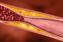
What happens to the spine with osteochondrosis? Why does pain occur? When playing sports, lifting weights, falling, a sick person develops microtraumas in the vertebra. With such frequent trauma to the spine, the intervertebral discs will lose their elasticity and elasticity, the distance between the vertebrae will decrease. This leads to pinching nerve endings, severe pain with osteochondrosis appear just because of this. And in this place, edema occurs in a short period of time. And this does not help to reduce pain in osteochondrosis, but only leads to its increase.
Doctors don't fully understand why some children get Panner's disease, but many believe the disease is hereditary. This is usually due to excessive and repetitive use of the elbow, which is under pressure and is forced during this period rapid growth bones. This overuse can result from participation in activities that involve throwing objects or applying heavy pressure to the joints, such as baseball and gymnastics. These mild and repetitive injuries cause the area to swell and irritate, which causes pain.
The location of the injured intervertebral discs also determines the place where pain is felt: the neck, shoulder, arm, and the chest may hurt. Pain in chest patients suffering from this disease are characterized as a stake in the chest.
The most common is chronic back pain.
Pain in osteochondrosis is the main symptom of the disease.
The main symptom of Panner's disease is Blunt pain around the outside of the elbow, near the humeral condyle. Typically, the pain gets worse when you do an activity, like throwing a ball, and it gets better when you rest. Children may also experience the following.
Insensitivity to touch swelling to fully expand the elbow inability to fully rotate the arm. Symptoms usually appear unexpectedly and may not be related to any specific event or trauma. The duration can be from several weeks to several months.
Sometimes there is sharp pain on the left, and the patient confuses the pain of a pinched nerve with heart pain. The patient often experiences pain in the right or left hypochondrium.
There is a feeling of numbness of the muscles and their strong tension.
If, as a result of osteochondrosis, they are compressed blood vessels, then the person has a headache, dizziness, tinnitus, nausea and vomiting.
With osteochondrosis lumbar spine patients experience pain in the lower back, often it is shooting and spreads to the leg and pelvis.
To determine if a child has Panner's disease, the doctor will first gather some information; to do this, he will ask about the age of the child, his activity, participation in sports events and his dominant group. He will press on and around the elbow to see if it causes pain. He will also ask the child to move his arm in certain directions to see how well he can move it and if the movement causes pain.
The doctor will likely give x-rays of the elbow to see if there are abnormalities and confirm the diagnosis. An x-ray allows the doctor to see the shape of the humeral condyle, which may be flat. In addition, they can show that the growth cartilage has an irregular or fragmented appearance and reveals areas where bone resorption occurs.
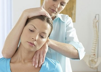
When the above symptoms appear, you need to seek help from a neurologist, traumatologist. The doctor will examine the patient, listen to complaints and, if he sees fit, prescribe X-rays, computed tomography or magnetic resonance imaging.
It is possible that some physicians use magnetic resonance imaging studies to more detailed analysis bones and assessment of the degree of swelling. Because the bones supply blood and repair themselves, most children with Panner's disease require very little treatment. The immediate goal is to relieve pain, and The best way to do this is to leave the injured elbow alone. This may include not participating in certain sports and sports until the elbow has healed.
Rest should help greatly relieve pain and swelling and gradually restore motion to the elbow. The doctor will monitor the recovery and determine when the child will start playing sports again. In general, children do not need to play sports for a long time.
Treatment Methods
This disease is very poorly treatable, so the patient must be patient. For the treatment of osteochondrosis, the following methods are used:
- medical method. Pain management involves the use of pain medications.
- Manual therapy helps a lot.
- Spinal traction is also used.
- Apart from manual therapy using acupuncture.
- Massage and therapeutic exercises help a lot.
- Some patients use traditional medicine in the treatment of this disease.
- If the patient has developed an intervertebral hernia, surgical treatment is used.
To prevent this disease, you must do the following:
The doctor may also recommend the following. Apply an ice pack or rub on the elbow to relieve pain and swelling, do physical therapy, especially if the child has difficulty folding and stretching the arms take an over-the-counter non-steroidal anti-inflammatory drug to relieve pain and swelling, such as acetaminophen or ibuprofen. Note. Children should not be prescribed aspirin for pain relief due to the risk of a very serious condition called Reye's syndrome.
If rest does not relieve pain, the doctor may recommend that the child wear a cast or splint around the entire arm to immobilize the elbow and allow it to heal. The child can use the cast for 3-4 weeks until the pain, swelling and tenderness subsides.
- Monitor the correct position of the body in childhood and take timely measures for violations of posture at this age.
- Study exercise. This will help build muscles, and they will already support the spine, and the posture will be correct.
- With this disease, diet is very important. You need to consume enough vitamins, calcium and magnesium. It's all in fish, cabbage, legumes, nuts and more.
- You don't have to be overweight.
- Equally load your hands when carrying heavy loads.
Being engaged in the treatment of osteochondrosis, one should not forget about folk methods. In combination with medical methods, physical exercises and all of the above methods, they give an excellent result. ethnoscience provides a lot of recipes based on medicinal herbs. Rubbing is done with various ointments, you can take baths using herbal infusions.
While recovery sometimes takes time, most children with Panner's disease are completely cured without any problems in the future. Over time, the child's bones mature, the brachial condyle grows back into its original shape, and pain and other symptoms usually disappear completely.
However, in some cases, children suffering from Panner's disease continue to have trouble expanding their arms fully even after treatment. Silva, Luis Claudio Lopez Correia da Roncati, Neymar Wanderlei Zoppa, Andre Luis do Valle de. Surgical treatment of osteochondrosis dissections in horses: a retrospective and critical analysis.
Get rid of dangerous papillomas FOREVER
A simple and proven way to get rid of papillomas and warts without dangerous consequences. Find out how >>
Headaches with cervical osteochondrosis
Headaches are a sign of many diseases, one of which is cervical osteochondrosis. In today's article, we will figure out why they arise and in what ways they can be effectively dealt with.
Articulation Arthroscopy Equine osteochondral fragment Osteochondrosis. Osteochondrosis is one of the major developmental orthopedic diseases affecting horses and is commonly defined as a failure in the process of endochondral ossification. Unfortunately, its mechanisms are not well defined, but genetic predisposition, imbalance or excess nutrition, endocrine factors, and biomechanical forces acting on the joints are thought to be involved in a multifactorial etiology. One of the manifestations of osteochondrosis is the dissection of osteochondrosis, when, after the interruption of the ossification process, the chondrocyte colonies are disrupted and the basal layer necrosis occurs, generating an area of fragility where biomechanical forces can lead to the separation of cartilage fragments or osteochondral.
![]()
Why does the head hurt with cervical osteochondrosis
Headaches in cervical osteochondrosis appear as a result of deformation of the intervertebral discs, which leads to a violation of the structures of the spine and compression of nerve endings and blood vessels. Doctors distinguish four main conditions that provoke the appearance of pain in the head with osteochondrosis of the neck.
Usually the lesion develops in the first year of life, but clinical signs may appear later or even go unnoticed. Within clinical signs the most common is joint effusion, which may or may not be associated with lameness. Although there are several forms of treatment, most authors recommend surgical removal with arthroscopy as it has a higher success rate with better functional and aesthetic results. There are diverging works in relation to the prognosis and when these animals should undergo surgery.
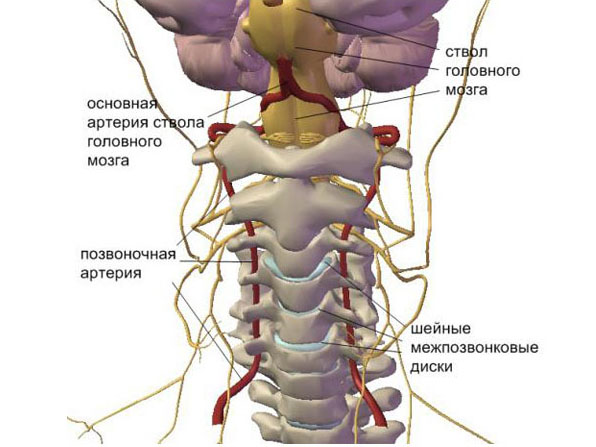
vertebral artery syndrome
Headaches in this case are long and throbbing, and are localized in the occipital region. After some time, pain can cover the parietal zone, and people also complain of pain in the temples. Analgesic drugs in this case almost do not help.
In order to answer these doubts and present the results obtained with surgical treatment in the medium and long term, this work was carried out. The lesions mainly affected animals under four years of age, during the pretreatment phase or during dressing, with most of them showing no clinical signs or lameness associated with joint effusion. Most of the lesions were located at the tibita-arsal joint, and in this joint the most affected site was the intermediate tibial crest.
The vertebral artery syndrome is caused by displacement of the vertebrae, herniated discs and protrusions. The disorder also causes a number of other additional symptoms, including dizziness and blackouts or blurry vision. This is due to poor blood circulation.
cervical migraine
When squeezing the nerve endings in the spine in the neck, pain occurs, passing into the right or left side head (depending on the location of the nerve). The pain is usually intense and lasts no more than 12 hours.
After the surgical procedure, 72% of the animals presented no clinical signs, and improvement was more significant in 3 and 4 year olds, and little improvement was achieved in lameness in animals over 6 years old. Surgical treatment of osteochondrosis dissections in horses: a retrospective study and critical analysis.
Arthroscopy End articular osteochondrosis. Osteochondrosis is the main development of an orthopedic disease affecting horses and is usually defined as a failure in endochondral ossification. Unfortunately, its mechanisms have not been determined, but it is believed to be a multifactorial etiology associated with genetic predisposition, nutritional imbalance or excess, endocrine factors, and biomechanical forces acting on the joints. Osteochondrosis dissecans, one of the representations of osteochondrosis, occurs after the cessation of the ossification process, disintegration of chondrocyte colonies and necrosis of the basal layer occurs, creating an area of weakness where biomechanical forces, when applied, can lead to separation of cartilage or osteochondral fragments.
Pinched nerve in the neck
Causes of headaches in cervical osteochondrosis may also lie in a pinched nerve. The patient in this case experiences almost the same as in the syndrome of the occipital artery. Headache with cervical osteochondrosis in this case smoothly passes into the parietal region and into the front of the head, creating pressure from the inside on the orbits. Pinched nerves in the back of the head as a result of displacement of the vertebrae, the appearance of bone growths or thinning of the interpeak disc.
Within clinical signs, total effusion is the most common, which may or may not be associated with lameness. Although there are various forms of treatment, most authors recommend surgical removal arthroscopically as this has a higher success rate with better functional and cosmetic results. There are conflicting studies regarding prognosis and the best time to refer an animal for surgery. To answer these questions and present the results obtained with surgical treatment in the medium and long term, this work was carried out.
Hypertensive syndrome
In this case, there is a bursting and pressing headache, the intensity of which increases when the head is turned. Pain usually does not last long - up to several hours. The occurrence of the syndrome is due to an increase in intracranial pressure as a result of squeezing of the vessels that pass through the narrowed canals in the vertebrae affected by osteochondrosis.
Most of the lesions were located in the articular joint, in the intermediate tibial crest. Postoperatively, 72% of the horses showed no clinical signs, and the improvement was more significant in animals at 3 and 4 years and little improvement in lameness in animals at six years. Effect of age and clinical manifestations in postoperative results surgical treatment osteochondritis with dissecans in horses. At the 12th World Equine Veterinary Association Congress in Hyderabad, arthroscopic approach to the volar notch of the distal interphalangeal joint through the flexor digitorum lamina in horses. Comparison of inflammatory response due to fluid or gas joint distension on arthroscopic examination in horses. Due to recent searches in my practice and mostly conversations with a few athletes different ages, levels of education and different genders about extremely painful syndrome in the leg, which hinders or even hinders learning, and, even worse, does not really effective treatment to Haglund's syndrome.
What can increase headaches in cervical osteochondrosis?
There are a number of factors that can provoke especially severe headaches in cervical osteochondrosis:
- sharp turns of the head;
- sedentary work;
- spinal injury;
- excess body weight;
- improper massage of the cervical region;
- uncomfortable bed;
- a diet that disrupts metabolic processes.
How to treat a headache with osteochondrosis of the cervical spine
The most important thing in the treatment of pain in the head with cervical osteochondrosis is the elimination of the root cause. To take off headache with cervical osteochondrosis, all your actions should be aimed at solving the following tasks:
A problem related to the mechanical impact of the posterior superior elevation of the posterior calcaneal tuberosity against the retrocalcaneal bursa and the Achilles tendon itself. This situation of local “impact” causes an inflammatory process. Since the patient is extremely uncomfortable and in the chronism of the picture, weakening and even local rupture of the tendon.
Trendy calcifications resulting from a non-specific chronic inflammatory process or secondary to mechanical hypersocialization of the region appear as true posterior calcaneus spurs. In addition to pain caused by internal inflammatory process, there is the possibility of friction and compression with the help of shoes, which aggravates the symptoms.
- relief of pain;
- normalization of blood circulation;
- restoration of normal neck mobility;
- relapse prevention.
Many people who do not know how to relieve a headache with cervical osteochondrosis begin to take painkillers, but they do not help, as usual. The fact is that the symptoms of the disease can be relieved only by the complex use of painkillers, antispasmodics and vasodilators. This helps to relax the muscles, stimulate the blood supply to the brain and relieve pain.
The patient during treatment requires peace and a comfortable place to sleep. It is recommended to buy an orthopedic pillow that will support the neck in a comfortable position. You can partially alleviate the condition with a massage of the collar area and head. for removal high blood pressure the doctor prescribes the appropriate drugs. Physiotherapeutic procedures such as:
- ozokerite;
- electrophoresis;
- magnetotherapy.