Antipyretics for children are prescribed by a pediatrician. But there are emergency situations for fever when the child needs to be given medicine immediately. Then the parents take responsibility and use antipyretic drugs. What is allowed to give to infants? How can you bring down the temperature in older children? What medicines are the safest?
The disease was described for the first time in 1965 by S. Hakim and R. D. Adams. . It is a variant of open hydrocephalus. This disease is characterized by slow expansion of the ventricular system with normal cerebrospinal fluid pressure. This leads to the development of a triad of symptoms (the Hakim-Adams triad). These include impaired walking, dementia, and urinary incontinence. .
Epidemiology. In the literature there are different data on this subject. The disease usually affects the elderly. The diagnosis is made in 0.41% of the population over 65 years of age, 0.4-6% in patients with dementia, and 15% with impaired walking. D. Jaraj et al. (2014) noted that normotensive hydrocephalus occurs in 0.2% of cases at the age of 70-79 years and in 5.9% - 80 years and older. However, the authors are of the opinion that the real figures are much higher. It is noteworthy that cases of diagnosing this disease are described even in childhood. The prevalence between men and women is the same. .
Etiology. Allocate primary and secondary normotensive hydrocephalus. In the first case, it is not possible to identify the causes of the development of the disease. Such patients account for half to one third of cases. Secondary hydrocephalus is a consequence of subarachnoid hemorrhage (30J, meningitis (15%), traumatic brain injury (10%), neurosurgical operations. .
Pathogenesis. At the heart of the disease is the formation of an imbalance between secretion and resorption of cerebrospinal fluid, impaired liquorodynamics. As a result, the volume of cerebrospinal fluid increases, and the volume of brain tissue decreases. This leads to irreversible ischemic and degenerative white and gray matter of the brain. This is characterized by the predominance of the frontal nature of neurological disorders. It is believed that this is due to the predominant expansion of the anterior horns of the lateral ventricles, which leads to compression of the deep sections. frontal lobes, anterior divisions corpus callosum, motor pathways connecting the cortex with lower limbs, dissociation of the basal nuclei from the frontal cortex, dysfunction of the frontal lobes, impaired sensorimotor integration. . In some cases, there is a decrease in blood flow to the brain. .
Clinical picture . The clinic of the disease is characterized by the gradual development of the classic triad of symptoms (walking disorder, dementia and urinary incontinence) over several months or years. However, after a traumatic brain injury or subarachnoid hemorrhage, symptoms may appear in the first days and weeks. .
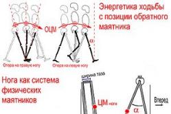
Walking disorders usually appear as the first signs of the disease. The gait slows down, then becomes unstable, falls are possible. Next, apraxia of walking manifests itself (uncertainty when standing and walking, difficulty initiating movement). While lying and sitting, the patient easily imitates the movements of walking, and in an upright position this is instantly lost. The strength in the limbs is not impaired. . Postural tremor, akinetic-rigid syndrome (the “freezing” phenomenon) may be detected. This brings the disease closer to the rigid form of Parkinson's disease (this diagnosis is often made initially). However, the examination does not reveal muscle rigidity. Sometimes there is a pseudobulbar syndrome. .
Cognitive impairments are characterized by a fronto-subcortical character; they usually develop against the background of already existing motor manifestations. . There are loss of short-term and long-term memory, disorientation in time. Patients have difficulty reporting their history. There are problems in planning, concentration, abstract thinking, violations of semantic memory. The emotional side becomes impoverished, apathy and complacency appear. Frequent phenomena of agnosia (violation various kinds perception: visual, auditory, tactile). The speed of mental processes and psychomotor reactions slows down. The degree of cognitive impairment varies. .
Violation of the function of the pelvic organs in the early stages is detected with active questioning - frequent urination and nocturia. Then there are imperative urges and then urinary incontinence. With the progression of cognitive impairment, patients lose their criticism of this problem and treat it indifferently. .
Diagnostics. Diagnosis is based on a triad of symptoms with ventricular dilatation and normal intracranial pressure.
Clinical and radiological criteria:
1 ) the presence of a complete or incomplete Hakim-Adams triad (gait disturbance, cognitive impairment, impaired control over the function of the pelvic organs, primarily urination)
2 ) the presence of a characteristic x-ray picture of NTG, which includes a combination of the following signs:
- expansion of the ventricles of the [brain] brain: Evans index more than 0.3 (30%) and expansion of the temporal horns of the lateral ventricles more than 2 mm (for reference: the Evans ventricular-hemispheric index is the ratio of the distance between the most distant points of the anterior horns of the lateral ventricles to the largest internal diameter of the skull)
- disproportionate expansion of the subarachnoid spaces (DESH-symptom): expansion of the cisterns of the base of the skull, lateral fissures of the brain in combination with compression of the interhemispheric fissure and parasagittal subarachnoid spaces in the parietal region.
In the diagnosis, a lumbar puncture is also used to determine the pressure of the cerebrospinal fluid. CSF pressure remains normal in this disease.
In addition, a TAP-TEST is carried out. It is also called the Miller-Fischer test, lumbar or spinal test. It is carried out as follows: a lumbar puncture is performed with the removal of 30-50 ml of cerebrospinal fluid. Before and after the test, a video recording of the gait is made. The test is considered positive if there is a significant improvement in gait or other symptoms after CSF evacuation. A positive test confirms the diagnosis of normotensive hydrocephalus. It has not yet been finally decided when and how to evaluate the results of this test. Basically, the assessment is made after 1 day. In normotensive hydrocephalus, gait and cognitive functions temporarily improve after this procedure. However, K. Kang et al. (2013) described a patient who did not respond after 1 day, but improved after 7 days. . The degree of improvement in the patient's condition after the test coincides with the effect of bypass surgery. Even a short-term decrease in the manifestations of at least one of the symptoms of the disease is considered a favorable prognostic sign. .
Treatment. The main method of treatment for these patients is bypass surgery with the imposition of a ventriculoperitoneal, ventriculoatrial or lumboperitoneal shunt. Positive results observed in 60% -66% -75% of patients. The prognosis of surgery worsens as the disease progresses. . It is preferable to use valve-controlled systems with an anti-siphon device and systems with a programmable variable pressure valve with the smallest possible step of opening pressure variation by design. .
IN postoperative period complex rehabilitation treatment is necessary.
Bibliography:
- Diseases nervous system: A guide for doctors: In 2 volumes - Vol. 1 / Ed. N. N. Yakhno, D. R. Shtulman. - 2nd ed. , revised and additional - M.: Medicine, 2001.
- https://laesus-de-liro.livejournal.com/285115. html
- Gusev E. N., Nikiforov A. S. Neurological symptoms, syndromes and diseases. - M.: GEOTAR-Media, 2006
- Moretti J.-L. “Assessment of alterations in cerebrospinal fluid kinetics. ” /In: Radionuclide Imaging of the Brain. Ed. by B. L. Holman. -New York etc.: Churchill Livingstone, 1985. -P. 185-223
- Adams R. D. , Fisher C. M. , Hakim S. et al. “Symptomatic occult hydrocephalus with “normal” cerebrospinal fluid pressure: a treatable syndrome.” //N. English J. Med. -1965. -Vol. 273.-P. 117-126
- Ketonen L. M. , Berg M. J. Clinical Neuroradiology. 100 Maxims. -London etc: Arnold, 1997
Normotensive hydrocephalus (NTH, Hakim-Adams syndrome) is a syndrome characterized by a combination of dementia, walking disorders and urinary incontinence with marked expansion of the ventricular system and normal CSF pressure. The prevalence of NTH is low, it is detected in 1-5% of patients with dementia. For the first time NTG as an independent disease was described by S. Hakim and R.D. Adams. In 1965, they published articles on "symptomatic occult chronic hydrocephalus in adults with normal fundus", or "hydrocephalus with normal CSF pressure." The authors paid special attention to the possible reversibility clinical manifestations this syndrome with adequate treatment.
Causes of normotensive hydrocephalus The development of the disease is based on an imbalance between the secretion and resorption of CSF, as well as a violation of liquorodynamics. NTG in adults can be induced by various causes:
- hemorrhage in the brain
- purulent-inflammatory process in the cranial cavity,
- perinatal lesions of the brain and meninges,
- volumetric intracranial formations,
- developmental anomalies of the brain common cause- Sylvian aqueduct atresia)
- undergone brain surgery.
Approximately 40-60% of cases in the anamnesis of patients with Hakim-Adams syndrome do not indicate any clear cause underlying the development of the disease (the so-called idiopathic IGT).
Clinical manifestations of Hakim-Adams syndrome
NTG is characterized by the gradual development of the Hakim-Adams triad - dementia, walking disorders and urinary incontinence. In most cases, gait disturbance is the first symptom, followed by dementia and later pelvic disorders follow. Possible fluctuation in the severity of symptoms, but this is not typical for IGT. The main complaint of patients with dacha pathology at the appointment with a neurologist is dizziness, which they describe as a feeling of instability during movement, sharp turns of the torso. In this case, the basis of dizziness is postural instability and dysbasia. characteristic of the disease. Walking disorders include elements of apraxia gait in the form of a shuffling gait with short, widely spaced steps and loss of balance control. With normotensive hydrocephalus, there are no changes in hand movements when walking, which distinguishes it from Parkinson's disease. In the early stages, with minimal support, there may be little change in gait in patients with normotensive hydrocephalus.
As the disease progresses, the step height decreases, it becomes difficult for patients to take their feet off the ground, difficulties appear in initiating the act of walking, turns are made in several stages. falls are frequent, postural instability may occur. It is necessary to carry out a differential diagnosis with Parkinson's disease and other extrapyramidal disorders. At the same time, patients with normotensive hydrocephalus can imitate the movements of the legs, which they must perform when walking, lying down or sitting. In idiopathic normotensive hydrocephalus, there is a relationship between the presence of arterial hypertension and the severity of clinical symptoms, especially walking disorders. Muscle tone in the legs, as a rule, is increased according to the plastic type or the type of resistance. In more severe cases of normotensive hydrocephalus, spasticity, hyperreflexia occur in the lower extremities, and pathological foot signs are detected. The presence of symptoms mainly in the legs in this pathology may be due to the fact that the motor pathways connecting the cerebral cortex with the lower extremities are located more medially - near the walls of the lateral ventricles, and the paths leading to the upper extremities are more lateral. Changes in gait in patients may also be due to dissociation of the basal ganglia from the frontal regions, dysfunction of the frontal cortex, and impaired sensorimotor integration.
Another important manifestation of Hakim-Adams syndrome is dementia. Patients are characterized by the presence of disorientation more in time than in place. It is often difficult for patients to state the history of their disease. Some may develop hallucinations, mania, depression. A characteristic symptom of normotensive hydrocephalus is also the development of emotional dullness. In general, cognitive impairments are manifested by a decrease in memory, a slowdown in the rate of mental processes and psychomotor reactions, a decrease in the ability to use acquired knowledge, and apathy, which is associated with dysfunction of the anterior parts of the brain and is characteristic of the so-called subcortical dementia.
Cognitive impairment in IGT is not the dominant syndrome, especially at the onset of the disease, when gnosis and other cortical functions are usually not impaired. Unlike Alzheimer's disease, memory impairment in normotensive hydrocephalus is not so pronounced and is caused by an ocular image of a decrease in the functional integration of the frontal lobes. Severe dementia in patients with normotensive hydrocephalus implies either an irreparable morphological defect (due to TBI, stroke, etc.) or the presence of concomitant Alzheimer's disease or vascular dementia. To detect cognitive disorders, especially in the early stages of the disease, neuropsychological methods sensitive to frontal disorders are used. The frontal nature of cognitive disorders in normotensive hydrocephalus may be due to the predominant expansion of the anterior horns of the lateral ventricles, accompanied by a more significant dysfunction of the deep compartments of the frontal lobes and anterior corpus callosum. In contrast to Alzheimer's disease, the cognitive defect in normotensive hydrocephalus develops more rapidly - within 3-12 months. The severity of cognitive disorders may decrease after administration of 20-50 ml of CSF (tap-test). It is believed that the basis of cognitive disorders in normotensive hydrocephalus is compression of the brain capillaries by increased intraparenchymal pressure, especially since, according to positron emission tomography, a diffuse decrease in metabolism is detected both in the cortical and 8 subcortical regions.
Already in the early stages, with active, purposeful questioning, it is possible to identify patients' complaints of frequent urination and nocturia. As the disease progresses, imperative urges and periodic urinary incontinence join. Patients stop feeling the urge to urinate and are indifferent to the fact of involuntary urination, which is typical for the frontal type of pelvic disorders. Fecal incontinence is rare, usually in patients at an advanced stage of the disease.
Results of additional surveys
It is important to note that during ophthalmoscopy, patients do not have congestion in the fundus. According to the EEG, in normotensive hydrocephalus, nonspecific changes in the bioelectrical activity of the brain are revealed, characterized by the predominance of slow frequency characteristics.
The results of CT and MRI make it possible to detect sharply dilated cerebral ventricles, while the cortical sulci remain within the normal range or slightly dilated. Using these techniques, other causes of hydrocephalus can be ruled out. The detection of small ischemic foci or areas of leukoaraiosis does not contradict the diagnosis of normotensive hydrocephalus, since a combination of Hakim-Adams syndrome and cerebrovascular insufficiency is possible. With normotensive hydrocephalus, the 111 ventricle, temporal and frontal horns of the lateral ventricles are especially significantly expanded, which leads to the appearance characteristic form ventricular system in the form of a "butterfly" on axial sections. The expansion of the anterior horns of the lateral ventricles in normotensive hydrocephalus reaches 30% or more of the diameter of the skull.
Surgical treatment of NTG syndrome
The basis of treatment is shunt operations of ventriculoperitoneal and lumboperitoneal shunting, in which a positive effect is achieved.
in 60% of patients. In advanced stages of the disease, when there are already irreversible changes in the brain, the prognosis of surgical treatment worsens. In some patients, among whom no
significant improvement after lumbar puncture, bypass surgery may also be effective. Complications after ventriculoperitoneal shunting in 30-40% of patients:
- subdural hematomas;
- liquor hypotension syndrome.
Mortality after ventriculoperitoneal shunting is about 6-7%. To prevent complications, an individual selection of a shunt is recommended.
Normotensive hydrocephalus conservative treatment
Purpose: to reduce the production of cerebrospinal fluid
Prescribed diacarb (acetazolamide) ...
Treatment of urination disorders is an extremely difficult task: the available drugs are ineffective.
Some patients, at least temporarily, are helped by teaching them to empty their bladders "on the clock".
Morgagni's syndrome occurs due to cerebral ischemia, which occurs with a sharp decrease in cardiac output. It occurs when there is an abnormal heart rhythm or heart rate.
Morgagni Adams Stokes attacks are often caused by atrioventricular block. An attack occurs when blockade occurs, followed by the development of sinus rhythm or supraventricular arrhythmia.
Causes, provoking diseases and factors
Attacks of the syndrome occur during such processes in the body:
- atrioventricular block;
- the transition of an incomplete atrioventricular blockade to a complete one;
- violation of the heart rhythm, which is accompanied by a decrease in myocardial contractility (with febrile, ventricular flutter, paroxysmal tachycardia, asystole);
- tachyarrhythmia and tachycardia with a heart rate of more than 200 beats;
- bradyarrhythmia and bradycardia at a heart rate of less than 30 beats.
The risk of developing the syndrome exists if the following conditions are present in the anamnesis:
- Chagas disease;
- inflammatory processes localized in the heart muscle, and which extend to the conduction system;
- diffuse proliferation of scar tissue, and subsequent heart damage in Lev-Legener disease, rheumatoid arthritis, Liebman-Sachs disease, systemic scleroderma;
- diseases with general neuromuscular changes (genetic diseases, myotonia);
- intoxication medicines(beta-blockers, calcium antagonists, cardiac glycosides, amiodarone, lidocaine);
- ischemia of the heart muscle in cardiomyopathies, myocardioclerosis, infarction;
- increased deposition of iron in hemochromatosis and hemosiderosis;
- systemic amyloidosis;
- functional conduction disturbances in the atrioventricular node.
Features of the clinical picture
The syndrome occurs in 25-60% of patients with complete atrioventricular block. The frequency and number of seizures varies in each clinical case. Attacks of Morgagni Edems Stokes can occur with a frequency of once every few years, and can occur several times within one day.
Sudden movements, sudden changes in body position, nervous overload, feelings, emotional stress can provoke an attack.
The attack is preceded by the following symptoms:
After some time (about 1 minute), the patient has an attack, and he loses consciousness. Fainting occurs when the heart rate is less than 30.
Fainting, often short-term, and does not last more than a few seconds. During this time, compensatory mechanisms are activated that allow the arrhythmia to be eliminated. After leaving this state, the patient has retrograde amnesia, and he does not remember what happened.
With an attack of Morgagni Adams-Stokes syndrome, the following symptoms are characteristic:
- pallor of the skin;
- swelling of the neck veins;
- blue lips and fingertips;
- spontaneous defecation and urination;
- cold sweat (sticky);
- weak muscle tone, cramps;
- inability to determine the pulse;
- reduced blood pressure;
- muffled and arrhythmic heart sounds;
- dilated pupils;
- rare and deep breathing.
Depending on the degree of intensity of the manifestation of symptoms, several forms of an attack are distinguished:
- Easy - there is no loss of consciousness, the patient experiences dizziness, sensitivity is disturbed, noise in the ears and head appears.
- Moderately severe - the patient loses consciousness, but there are no signs such as voluntary urination and defecation, convulsions are also not observed.
- Severe - the whole symptom complex is present.
Emergency First Aid
With an attack of the Morgagni-Adams-Stokes syndrome, the patient needs urgent health care, on which the duration of the attack itself and the life of the patient will depend.
First of all, it is made mechanical defibrillation, also referred to as a precordial beat. It is necessary to strike with a fist in chest, namely in its lower part. You can not make a blow to the heart. After mechanical defibrillation, the heart reflexively begins to contract.
If there is no effect, electrical defibrillation is performed. To do this, electrodes are placed on the patient's chest and a shock is made with a current discharge. After that, the correct rhythm of the heartbeat should return.
In the absence of breathing, artificial ventilation of the lungs is performed. To do this, air is blown into the patient's mouth using a special apparatus, or according to the mouth-to-mouth method.
Cardiac arrest is an indication for an injection of Adrenaline (intracardiac) or Atropine (subcutaneous).
If the patient retains consciousness, then he needs to give the drug Isadrin under the tongue (the effect is similar to Adrenaline, Ephedrine, Norepinephrine, but there is no increase blood pressure).
The patient must be taken to the intensive care unit of the hospital. In the hospital, emergency care is accompanied by monitoring on an ECG machine. Atropine sulfate and Ephedrine are administered subcutaneously several times a day to the patient, Isadrin is given under the tongue. If necessary, electrical stimulation is carried out.
Establishing diagnosis
Loss of consciousness is possible with various diseases. Therefore, when conducting a diagnosis, the Adams-Stokes-Morgagni syndrome must be differentiated from the following conditions:
To determine the syndrome, the following diagnostic methods are used:
- electrocardiogram (ECG) in dynamics;
- monitoring of the cardiogram on the Holter apparatus (allows you to identify temporary changes, a combination of flutter and atrial fibrillation);
- long-term monitoring of the electroencephalogram;
- contrast coronography of vessels;
- myocardial biopsy.
Treatment of the syndrome
Starting treatment means emergency care at the onset. This is followed by therapy, the purpose of which is the prevention of recurrence of attacks of Morgagni Adams-Stokes syndrome. Therapeutic measures are carried out in the cardiology department.
Initially, the causes of seizures are identified, a detailed examination of the heart is carried out, the diagnosis is clarified and a set of therapeutic measures is prescribed. Such methods of therapy of the syndrome are used.
Medical treatment
After the patient is admitted to the intensive care unit, treatment with medications is performed. Droppers are used with the introduction of Ephedrine, Orciprenaline. Every 4 hours the patient is given Isadrin. Injections of Ephedrine, Atropine are made.
Inflammatory processes are removed with the help of corticosteroids. Since bradycardia is accompanied by tissue acidosis and hyperkalemia, it is necessary to take diuretics, an alkaline solution. This helps to remove potassium from the body and normalize blood pressure.
After the attack stops, preventive therapy is prescribed with the use of antiarrhythmic drugs, and therapeutic measures are directed to get rid of the main cause of the syndrome (ischemia, intoxication, inflammatory process).
Surgery
If there is a risk sudden stop heart and recurrence of seizures, then a necessary measure is the implantation of a pacemaker. It is possible to use two types of pacemakers: with a complete blockade - a device that provides constant stimulation of the heart, with an incomplete blockade - a device that works when violations occur.
During surgical operation the electrode is inserted through the vein and fixed in the right ventricle of the heart. The body of the stimulator is fixed in the rectus abdominis muscle (in men) or in the retromammary space (in women).
The pacemaker should be checked for performance every 3-4 months.
Prevention of seizures and relapses
Application preventive measures possible with attacks that are caused by paroxysms of tachyarrhythmia or tachycardia. In this case, patients are prescribed various antiarrhythmic drugs.
You should also exclude the factors that lead to the onset of an attack - sudden movements, sudden changes in body position, experiences, nervous overload, emotional stress, intoxication.
With complete atrioventricular blockade, the main preventive method is the installation of a pacemaker.
What is the risk?
The severity of the consequences directly depends on the frequency of seizures and their duration. Frequent and prolonged hypoxia of the brain entails a negative prognosis of the disease.
Hypoxia lasting more than 4 minutes brings irreversible brain damage. Lack of resuscitation measures ( indirect massage heart, artificial respiration) can lead to the cessation of cardiac activity, the disappearance of bioelectrical activity and death.
When performing a surgical operation, the prognosis is positive. Implantation of a pacemaker allows you to restore the quality of life, working capacity and health of the patient.
This section was created to take care of those who need a qualified specialist, without disturbing the usual rhythm of their own lives.
Biology and medicine
Normal pressure hydrocephalus (Hakim-Adams syndrome)
Hakim-Adams syndrome (normotensive hydrocephalus) is a condition that is often talked about but difficult to diagnose. Normal pressure hydrocephalus is a syndrome with characteristic clinical and pathophysiological features, as well as typical changes on CT or MRI. The classic triad is a gait disorder as a result of ataxia or apraxia (Combination of walking disorders by the type of frontal dysbasia (walking apraxia) with dementia and urinary incontinence).
CT or MRI reveals enlargement of the lateral ventricles (hydrocephalus) with little or no atrophy of the cerebral cortex. Hydrocephalus - communicating, we pass the Sylvian aqueduct (Fig. 367.3). CSF pressure during lumbar puncture corresponds to upper bound norms. The content of protein and glucose in the CSF is not changed. It is believed that normotensive hydrocephalus develops as a result of impaired CSF flow along the upper lateral surface of the hemispheres and CSF absorption in venous system. Since the disease develops slowly, the lateral ventricles dilate, but CSF pressure rises slightly.
The cause of the clinical manifestations of normotensive hydrocephalus is unknown. Perhaps they are due to stretching of the fibers of the radiant crown of the brain. Sometimes normotensive hydrocephalus develops after diseases leading to the formation of adhesions on the membranes of the basal surface of the brain, such as meningitis, subarachnoid hemorrhage, or traumatic brain injury. However, in most cases, normotensive hydrocephalus develops for no apparent reason.
In contrast to Alzheimer's disease, normotensive hydrocephalus develops early gait disturbances, and CT or MRI does not reveal cortical atrophy. Attempts have been made to find research methods to confirm the diagnosis of normotensive hydrocephalus and predict the outcome of shunting. Among them are cisternography (allows you to diagnose a slowdown in CSF absorption on the upper lateral surface of the hemispheres) and various liquorodynamic studies. None of these studies are specific enough to be used in daily practice.
In some cases, gait and cognitive functions temporarily improve after CSF extraction, however, this cannot serve as a criterion for the effectiveness of the upcoming bypass. According to one study, no more than 1-2% of cases of dementia are caused by normotensive hydrocephalus.
Sometimes this disease is misdiagnosed as Alzheimer's disease, since the latter can be accompanied by gait disturbance, and cortical atrophy at an early stage of the disease is not always visible on CT or MRI. characteristic feature Alzheimer's disease in this case may be atrophy of the hippocampus on MRI.
Shunting with proven normotensive hydrocephalus is effective in 30-50% of cases. After surgery, sometimes gait is restored better than memory. Often the improvement is short-lived. Patients for surgery are carefully selected, since it is often accompanied by subdural hematoma and secondary infection.
The clinical triad is characteristic: memory loss, gait disturbance and urinary incontinence. The first symptom is often impaired gait, and the dementia is usually mild. CT and MRI of the brain reveal dilatation of the lateral ventricles, but there is no or minimal cortical atrophy. On lumbar puncture, CSF pressure is normal or slightly elevated, and CSF composition is unchanged.
Normotensive hydrocephalus can be either idiopathic or due to previous meningitis or subarachnoid hemorrhage (due to ruptured aneurysm or traumatic brain injury).
A likely pathogenetic mechanism is blockade of CSF outflow along the upper lateral surface of the brain, as well as difficulty in the absorption of CSF into the blood, which leads to stretching and twisting of the pathways in the radiant crown. Sometimes ventricular shunting helps.
Differential diagnosis of normotensive hydrocephalus with Alzheimer's disease is difficult.
Links:
Random drawing
Attention! Information on the site
intended solely for educational
Normotensive hydrocephalus, or Hakim-Adams disease
1. Definition 2. Etiology and pathogenesis 3. Symptoms of idiopathic hydrocephalus 4. Gait disorders 5. Dementia 6. About pelvic disorders 7. Diagnosis 8. Treatment 9. Complications and prognosis
When doctors talk about hydrocephalus, almost always the leading mechanism for its development is an increase in intracranial pressure (increased ICP syndrome), or intracranial hypertension. It is she who is to blame for the appearance of such symptoms as a progressive decrease in vision, bursting headaches and vomiting. In the event of a sudden obstruction to the flow of CSF, occlusive hydrocephalus occurs, and then the brain may begin to swell and be infringed.
These types of hydrocephalus are described in detail here. But it turns out that there is also a form of the disease that manifests itself with completely different symptoms and often goes unnoticed. We are talking about the so-called normotensive hydrocephalus, or Hakim-Adams syndrome. What is this pathology and can this disease be cured?
Definition
This is the name of the condition in which the pressure of the cerebrospinal fluid almost always does not exceed the permissible values, but at the same time there is dropsy of the ventricular system of the brain. Since the pressure is within the normal range, the symptoms of such a pathological condition should be completely different. And this is true: it was not until 1965 that the first generalized data were published in The New England Journal of Medicine. The authors of the material were R. Adams, S. Hakim and C. Fisher. Since no name has yet been coined for this condition, they described "occult hydrocephalus in adults with a chronic course without changes in the fundus and with normal CSF pressure."
Thus, a hydrocephalus syndrome was considered, devoid of its basic and supporting diagnostic features. Everyone knows that at the first "classic" complaints associated with a presumptive increase in ICP, the doctor assesses the condition of the patient's fundus. In the event that there are no changes on it, then the diagnosis of hydrocephalus is considered doubtful. And here a mysterious condition was described, in which not only was the fundus normal, but no characteristic complaints were observed - the flow was hidden.
Nevertheless, this condition turned out to have its own “markers” - dementia, gait disorders and urinary incontinence. If these symptoms are accompanied by a pronounced expansion of the ventricles of the brain, then the diagnosis of normotensive hydrocephalus is made. According to the ICD-10, he was assigned the code G 91.2, or “normal pressure hydrocephalus”.
The great difficulty in recognizing this condition is that such signs are often found in elderly patients with various forms of dementia, and no one knew about the presence of hydrocephalus when the mentioned symptoms were detected. The name “Hakim-Adams syndrome” was given to the disease, while for some reason the name of C. Fisher was not indicated, which was between R. Adams and S. Hakim in the list of authors.
Etiology and pathogenesis
It should not be thought that this is a common disease and normotensive hydrocephalus should be suspected in every elderly patient with pelvic disorders: its prevalence does not exceed 4% among people suffering from various forms of dementia. Complicating the situation is the fact that the diagnosis of dementia itself has different criteria, and so far there is no single approach among scientists from different countries.
What is the cause of NH (normotensive hydrocephalus)? In about half of all cases, it was possible to prove that the patient had a history of:
- hemorrhages in the brain (both intraventricular and subarachnoid);
- various craniocerebral injuries;
- meningitis;
- perinatal damage to the central nervous system;
- volumetric formations in the cranial cavity (from castes and aneurysms to tumors);
- congenital anomalies of the cerebrospinal fluid system - atresia, or underdevelopment of the Sylvian aqueduct;
- operative neurosurgical interventions.
It is clear from this list that nothing specific, leading to pathology, was identified - the list turned out to be too “variegated”, and it must be taken into account that the doctors were specifically looking for the cause. In 50% of cases, no possible factors were found at all. Given this circumstance, they agreed to call this condition “idiopathic hydrocephalus”. The situation was saved, and the cause of the Hakim-Adams syndrome could "officially" remain unknown.
Interestingly, without an increase in ICP, it is impossible to "inflate" the ventricles. How does the paradox arise? normal level ICP with dilated ventricles? It turned out that episodes of ICP rise exist, but they occur at the same time as Prinzmetal's spontaneous angina - at night, during REM sleep. The vessels dilate, hyperemia of the brain occurs. The outflow of CSF is also disturbed, which is functional in nature, associated with a change in pressure indicators in different parts of the CSF system of the brain. IN daytime, as well as in the phase of slow sleep, this violation does not occur.
Since the episodes of the rise in ICP are temporary, and not permanent, then the symptoms of "stagnation" are not determined - it simply does not have time to develop.
How is this disease detected?
Symptoms of idiopathic hydrocephalus
Fortunately, the clinical manifestations of the disease are much more definite than possible reasons. This is the “Hakim-Adams triad”: first, gait is disturbed, later dementia progresses and its diagnosis is made, and only then pelvic disorders occur, among which urinary incontinence comes to the fore. Symptoms can be expressed with varying intensity, but variability is quite acceptable. What are the characteristics of each "member of the triad"?
Gait disorders
The gait of a patient with NG is uncertain: he overcomes turns poorly, moves in small steps, hardly maintains balance and tries not to raise his legs above the floor plane. Such a gait is also called "stuck", or "magnetic". Only the movements of the legs are striking, with the hands of the patients everything is in order.
In patients, step height gradually decreases, it becomes difficult for them to climb stairs. It is just as difficult to start moving, and in order to turn, you need to perform several “preparatory” actions. In this case, the patient often falls. It is noteworthy that the patient can perform stereotypical "walking" movements with his legs, even while lying in bed or sitting.
Muscle tone does not determine the development of gait disturbance and its skills (gait apraxia). It can be diffusely reduced and remain physiological or elevated in a pyramidal or spastic type.
A characteristic feature that makes it possible to determine the Hakim-Adams syndrome is a sharp improvement in the patient's gait after he is given a lumbar puncture and a rather large (20–40 ml) amount of CSF is removed. This test is called "tap-test". As noted, the removal of cerebrospinal fluid significantly improves the ability to keep balance, and at the same time walking is restored.
Some scientists believe that the functions of the hands remain normal because their motor fibers go further from the walls of the lateral ventricles of the brain than the fibers of the legs, and undergo minimal morphological changes.
dementia
Dementia, or a violation of higher cortical functions, is fully manifested in this disease, usually after gait disorders. But often it precedes them, and in a non-specific form: memory decreases and the rate of mental reaction slows down. Apathy sets in. In the future, complacency arises, aspontaneity appears, or the impulse to any kind of activity decreases. Disorientation in time is observed, and in some patients cognitive disorders transform into psychotic ones: hallucinations, a manic state appear, and even consciousness is disturbed by the type of delirium.
Others and relatives notice symptoms of "emotional atrophy": patients stop showing any feelings. In a severe course of the disease, drowsiness, a soporous state, and even akinetic mutism may develop. All this leads to the occurrence of bedsores, the addition of a secondary infection and the death of the patient.
There is also a tendency to the manifestation of the "frontal" psyche. The “frontal” nature of the disorders consists in a decrease in self-criticism, the appearance of foolishness, a tendency to flat and “greasy” jokes, as well as a violation of the sequence of actions. So, the patient can first urinate, and then unbutton his pants. Many researchers believe that this is due to the predominant lesion of the anterior horns in the ventricles. As a result, the work of the frontal lobes in depth is disrupted, their associative and commissural connections between themselves and with the corpus callosum are depleted.
Cognitive impairment in Hakim-Adams disease occurs faster than in "classic" dementia, such as Alzheimer's syndrome. So, after 9-12 months from the onset of cognitive disorders, you can get a patient who will constantly need the help of others.
About pelvic disorders
If you carefully question the patient with a complaint of difficulty in gait, then already at the beginning of the disease, phenomena such as nocturia (the predominance of nighttime urination over daytime), as well as dysuric disorders, which are manifested by frequent urination, can be identified. Then there are imperative urges: the patient urgently needs to go to the toilet, and if there is no suitable place, the urine is simply not retained.
In the future, with the progression of the "frontal psyche", the patient becomes indifferent to both the imperative urges themselves, and the urinary incontinence that develops later, even without any urges. As a rule, patients with normotensive hydrocephalus do not suffer from fecal incontinence. However, this may occur at a later stage, in bedridden patients.
When conducting a "tap-test", it is possible to restore the function of the pelvic organs for a while: the patient begins to hold urine. Such a reaction does not occur in other types of PTO disorders (function of the pelvic organs).
Can any symptoms occur in patients other than the Hakim-Adams triad? The answer is yes, but taken separately, they have no independent diagnostic value, since they speak of a general involution of the central nervous system. This may be pseudobulbar syndrome, grasping and proboscis reflex, some types of tremor.
Diagnostics
It was noted above that the difficulty in detecting the disease is due to the wide spread of the main symptoms in the elderly in general, as well as the insufficient level of medical care for this category of the population.
It is useless to carry out all kinds of routine analyzes, their information content is zero. The main diagnostic criteria are:
- the presence of a supporting "triad";
- detection of signs of internal hydrocephalus on MRI with an increase in the lateral ventricles (their anterior horns can be 1/3 of the diameter of the skull, and the picture resembles a "butterfly").
External hydrocephalus, especially isolated, with the preservation of the normal size of the lateral ventricles, does not allow the diagnosis of "Hakim-Adams syndrome", as well as the presence of thinned and atrophic sulci and convolutions. In normotensive hydrocephalus, the cortex is practically unchanged. On the other hand, in older patients one can hardly see the "ideal" cortex. Usually there are signs of ischemia, the presence of small foci of gliosis, leukoaraiosis. All this indicates chronic cerebrovascular diseases and does not contradict the diagnosis of Hakim-Adams syndrome.
During the MRI, it is necessary to carefully assess the condition of the Sylvian aqueduct and the outflow of cerebrospinal fluid through it. This will allow you to decide on further treatment tactics, if bypass surgery is needed. Therefore, it is preferable to do an MRI rather than a CT scan, since it is on an MRI that the structures of the brain are clearly visible.
"Normal" refers to the absence of congestion and disc edema optic nerves. Everything else may be present according to age and concomitant diseases.
After that, they begin to conduct a lumbar puncture - the main test. Normal cerebrospinal fluid pressure is detected, which does not exceed 180–200 mm of water. Art. It is known that this pressure fluctuates normally and is associated with the pulse, respiration and blood pressure level - the amplitude of fluctuations is usually 10% of the CSF pressure level (no more than 20 mm of water column). In normotensive hydrocephalus, the amplitude of such fluctuations is much higher.
An important method of confirming the diagnosis is a “fantastic” study for domestic pensioners - night monitoring of intracranial pressure, carried out in conjunction with polysomnography. If its increase coincides with the phase of REM sleep and regression during wakefulness, it is possible to make a diagnosis of Hakim-Adams syndrome with full right. Before this study, transcranial dopplerography should be performed. It will allow to evaluate the relationship between the dynamics of CSF pressure pulsation and blood flow in the vessels of the brain. This is necessary for the correct interpretation of the results.
The diagnostics is completed by the implementation of a tap-test and other methods. In particular, the dynamics of the decrease in the level of cerebrospinal fluid pressure after the introduction of endolumbar saline is studied.
In addition, to assess the degree of dementia, it is necessary to use special tests, scales and neuropsychological surveys that are “sensitive” specifically to the frontal psyche. If you use the usual short scale, you can not detect frontal disorders, and this is a standard error of neurologists. But there is simply no single technique that allows 100% to diagnose the frontal psyche.
Treatment
Conservative treatment of Hakim-Adams syndrome is carried out by prescribing Diakarba (acetazolamide) and digoxin cardiac glycoside. But the proven effectiveness of this type of therapy has not been identified. The most difficult task is the treatment of urinary disorders in patients. For this, the use of anticholinergic drugs is recommended in order to at least develop in patients the skill of emptying Bladder at the right time.
But the possibilities of conservative treatment of the disease are limited: non-surgical methods do not prolong life and do not lead to an improvement in its quality. Therefore, shunting is the world standard in the treatment of NG. After all, it is the decrease in CSF pressure, despite its normal numbers, that leads to a sharp decrease in symptoms (gait and cognitive functions improve).
Most often, ventriculoperitoneal shunting is used, with persistent positive dynamics of 60% or more, especially with early surgery after several months of illness.
Complications and prognosis
In the event that timely surgical intervention is performed, the risk of complications in elderly patients is 30%. This is a high figure, but, as a rule, there is a postoperative hematoma, "sticking" of the ventricles due to a sharp drop in pressure and CSF hypotension. To eliminate these consequences, it is required to choose the right shunt and calculate the optimal pressure.
Shunting leads to the fact that in 50-70% of all cases the condition stabilizes, the symptoms regress, and the patient is no longer in danger of disability, especially with an early onset of the disease. The prognosis for normotensive hydrocephalus, which is treated conservatively, is doubtful, and even unfavorable: within a year, the development of pelvic disorders and the progression of dementia are possible.
In conclusion, it must be said that such a pathology as the Hakim-Adams syndrome is a “litmus test” of the state of health care in the country and attitudes towards old age. If the problem of an elderly person is treated with attention, doctors do not just diagnose dementia, but look for the cause - this is a step towards recovery.
Comments (0)
Write a comment
Diseases
Would you like to proceed to the next article "Hydrocephalus in adults: causes, varieties, clinic, and principles of treatment"?
Copying of materials is possible only with an active link to the source.
Morgagni-Adams-Stokes syndrome: causes, signs, diagnosis, help and treatment
Morgagni-Adams-Stokes syndrome (Morgani-Adams-Stokes, MAS, MES) is a sudden disturbance of the heart rhythm, which leads to its stop, disruption of blood transport to the organs and, above all, to the brain. Pathology is characterized sudden fainting, entailing disruption of the central nervous system, which manifests itself within seconds after cardiac arrest. The result of an attack of MAS syndrome can be clinical death.
According to statistics, up to 70% of patients with permanent complete atrioventricular block have manifestations of the MAC syndrome. In pediatric practice, this syndrome is usually observed in children with atrioventricular blockade of 2-3 degrees and sick sinus syndrome.
The severity of manifestations of the MAS syndrome and the frequency of an attack depend on its cause, the initial state of the heart and blood vessels, and metabolic changes in the myocardium. In some cases, seizures can be short-lived and go away on their own, but severe arrhythmias and cardiac arrest require emergency resuscitation, so these patients need increased attention from cardiologists.
Causes of MAC Syndrome
The conduction system of the heart is represented by nerve fibers, along which impulses move in a strictly specified direction - from the atria to the ventricles. This ensures the synchronous operation of all chambers of the heart. If there are obstacles in the myocardium (scars, for example), additional conduction bundles formed in utero, the mechanisms of contractility are violated, and prerequisites for arrhythmia appear.
example of MAC syndrome due to bradycardia
In children, among the causes of conduction disorders are birth defects, violations of the intrauterine laying of the conducting system, in adults - an acquired pathology (diffuse or focal cardiosclerosis, electrolyte disturbances, intoxication).
An attack of MAS syndrome is usually provoked by various factors, including:
- Complete AV block, when the impulse from the atria does not reach the ventricles;
- Transformation of an incomplete blockade into a complete one;
- Paroxysmal tachycardia, ventricular fibrillation, when the contractility of the heart muscle drops sharply;
- Tachycardia over 200 and bradycardia below 30 beats per minute.
It is clear that such severe arrhythmias do not occur by themselves, they need a substrate that appears when myocardium is damaged due to coronary disease, after a heart attack, inflammatory processes (myocarditis). Intoxication may play a role medicines from the group of beta-blockers, cardiac glycosides. Patients with rheumatic diseases (systemic scleroderma, rheumatoid arthritis) when involvement of the heart with inflammation and sclerosis is likely.
Depending on the prevailing symptoms, it is customary to distinguish several variants of the course of the MAS syndrome:
- Tachyarrhythmic, when the heart rate reaches, the function of blood ejection into the aorta suffers sharply, the organs experience hypoxia and ischemia.
- Bradyarrhythmic form - the pulse decreases for up to a minute, and the cause is usually a complete atrioventricular block, weakness of the sinus node and its complete stop.
- Mixed type with alternating paroxysms of asystole and tachycardia.
Features of symptoms
In MAS syndrome, attacks occur suddenly, and they can be preceded by stress, severe nervous tension, fear, excessive physical activity. A sharp change in body position, when the patient quickly gets up, can also contribute to the manifestation of heart pathology.
Usually, in the midst of full health, a characteristic symptom complex of MAS appears, including cardiac disorders and brain dysfunction with loss of consciousness, convulsions, involuntary defecation and urination.
The main symptom of the disease is loss of consciousness, but before it the patient feels some changes, which he can later talk about. Darkening of the eyes, severe weakness, dizziness and noise in the head speak of an approaching fainting spell. A cold clammy sweat appears on the forehead, a feeling of nausea or dizziness appears, a feeling of palpitation or fading in the chest may appear.
Seconds after the paroxysm of arrhythmia, the patient loses consciousness, and the surrounding people fix the signs of the disease:
- Lack of consciousness;
- The skin turns pale sharply, cyanosis is possible;
- Breathing is shallow and may stop completely;
- Blood pressure drops;
- The pulse is thready and often not palpable at all;
- Convulsive twitching of muscles is possible;
- Involuntary emptying of the bladder and rectum.
If the attack lasts for a short time, and the rhythmic contractions of the heart are restored by themselves, then consciousness returns, but the patient does not remember what exactly happened to him. With prolonged asystole, lasting up to five or more minutes, clinical death occurs, acute cerebral ischemia, and emergency measures are no longer enough.
The disease can proceed without loss of consciousness. This is typical of young patients in whom the vascular walls of the brain and coronary arteries are not damaged, and the tissues are relatively resistant to hypoxia. The syndrome is manifested by severe weakness, nausea, dizziness with preservation of consciousness.
Elderly patients with atherosclerosis of the cerebral arteries have a worse prognosis, and their attacks are more severe, with a rapid increase in symptoms and a high risk of clinical death, when there is no heartbeat and breathing, pulse and pressure are not detected, pupils are dilated and do not respond to light.
How to make the correct diagnosis?
In the diagnosis of MAC syndrome, the main importance is given to electrocardiographic techniques - ECG at rest, daily monitoring. To clarify the nature of the pathology of the heart, it can be prescribed ultrasonography, coronary angiography. Of no small importance is auscultation, when the doctor can listen to peculiar noises, amplification of the first tone, the so-called three-term rhythm, etc., but all auscultatory signs necessarily correlate with electrocardiography data.
Since Morgagni-Adams-Stokes syndrome is a consequence of various kinds of conduction disorders, electrocardiographic diagnostic criteria as such, it does not have, and the phenomena on the ECG are associated with the type of arrhythmia that is provoked in a particular patient.
In case of violation of conduction from the atrial node on the ECG, first of all, the duration of the PQ interval is evaluated, which reflects the time of passage of the impulse through the conduction system from the sinus node to the ventricles of the heart.
With the first degree of blockade, this interval exceeds 0.2 seconds, with the second degree, the interval gradually lengthens or exceeds the norm in all cardiac complexes, while QRST periodically drops out, which indicates that the next impulse simply did not reach the ventricular myocardium. In the third, most severe degree of blockade, the atria and ventricles contract on their own, the number of ventricular complexes does not correspond to the P waves, that is, the impulses from the sinus node do not reach the endpoint in the conductive fibers of the ventricles.
variety of arrhythmias causing MAC syndrome
Tachycardia and bradycardia are set based on the count of the number of heart contractions, and ventricular fibrillation is accompanied by a complete absence of normal teeth, intervals and the ventricular complex on the ECG.
Treatment of the MAC syndrome
Since MAC syndrome is manifested by sudden attacks of loss of consciousness and brain dysfunction, the patient may need emergency care. It often happens that a person falls and loses consciousness in a public place or at home in the presence of relatives, then the latter should immediately call an ambulance team and try to provide first aid.
Of course, others may be confused, not know where to start resuscitation, how to carry it out correctly, but in the event of a sudden cardiac arrest, minutes count, and the patient may die right in front of eyewitnesses, so in such cases it is better to do at least something to save a person’s life, because procrastination and inaction cost lives.
- Precordial stroke.
- Indirect cardiac massage.
- Artificial respiration.
Most of us have heard of tricks in one way or another. cardiopulmonary resuscitation but not everyone has these skills. When there is no confidence in your skills, you can limit yourself to pressing on the chest (about 2 times per second) until the ambulance arrives. If the resuscitator has already encountered such manipulations and knows how to do them correctly, then for every 30 clicks he performs 2 exhalations according to the “mouth to mouth” principle.
A precordial punch is an intense push with the fist to the region of the lower third of the sternum, which often helps to restore the electrical activity of the heart. A person who has never done this should be careful, because a strong blow from a fist, especially a man's, can cause a fracture of the ribs and bruises of the soft tissues. In addition, this technique is not recommended for young children.
Chest compressions and artificial respiration can be done alone or with a partner, the second is easier and more effective. In the first case, there are 2 exhalations for 30 compressions, in the second - one exhalation of pressure on the chest.
The ambulance team in case of cardiac arrest will continue emergency care, supplementing it with medical support. To restore the heart rhythm, pacing is performed, and if it is impossible to implement it, adrenaline is injected intracardiac or into the trachea.
In order to restore the conduction of impulses from the atria to the ventricles, atropine is indicated intravenously or subcutaneously, the introduction of which is repeated every 1-2 hours due to the short duration of the drug. As the patient's condition improves, he is given isadrin under the tongue and transported to a cardiological hospital. If atropine and isadrine do not have the expected result, then orciprenaline or ephedrine is administered intravenously under strict control of the heart rate.
In the bradyarrhythmic form of MAS, treatment includes temporary pacing and the administration of atropine, in the absence of which, aminophylline is indicated. If after these drugs the result is negative, dopamine, adrenaline are administered. After stabilization of the patient's condition, the issue of permanent pacing is considered.
The tachyarrhythmic form requires the elimination of ventricular fibrillation through electrical impulse therapy. If tachycardia is associated with the presence of additional pathways in the myocardium, then the patient will need further surgery to transect them. With ventricular variants of tachycardia, a pacemaker-cardioverter is installed.
In order to avoid attacks of cardiac arrest, patients with MAC syndrome are prescribed prophylactic antiarrhythmic therapy, including drugs such as flecainide, propranolol, verapamil, amiodarone, etc. (appointed by a cardiologist!).
If conservative treatment with antiarrhythmics does not work, there is a high risk of complete atrioventricular block and cardiac arrest, then pacing is indicated with the installation of a special device that supports the work of the heart and at the right time gives it the necessary impulse to contractions.
The pacemaker can work continuously or "on demand", and its type is selected individually based on the characteristics of the course of the disease. With a complete blockade of the conduction of impulses from the atria to the ventricles, it is advisable to use a pacemaker that works continuously, and with a relative safety of the automatism of the heart, an apparatus operating in the “on demand” mode can be recommended.
Morgagni-Adams-Stokes syndrome is a dangerous pathology. Sudden attacks of loss of consciousness and the likelihood of clinical death require timely diagnosis, treatment and observation. Patients with MAS syndrome should regularly visit a cardiologist and follow all his recommendations. The prognosis depends on the type of arrhythmia and the frequency of cardiac arrest, and timely implantation of a pacemaker significantly improves it and allows the patient to prolong life and get rid of asystole attacks.
Normotensive hydrocephalus (G91.0) is hydrocephalus that develops as a result of traumatic brain injury, subarachnoid hemorrhage, purulent meningitis, and there is no intracranial hypertension.
Normotensive Hakim-Adams hydrocephalus is characterized by a triad of symptoms: gait disturbance due to ataxia or frontal dysbasia due to apraxia, dementia, urinary disorders.
Normal pressure hydrocephalus occurs in 0.4-6% of patients with dementia. Prevalence: 2.5-5 per 100 thousand people. In 50% of cases, it is combined with anomalies in the development of the ventricular system.
Symptoms of normotensive hydrocephalus
Initially, there is a violation of walking (“legs stick to the floor”, slowing down of walking, difficulty in starting to move, imbalance / falling) for several months, years. Cognitive impairments (difficulty in thinking, memory loss) appear within 6-12 months. In the early period of the disease, urination disorders are added.
An objective examination of the patient can reveal ataxia, apraxia of walking (85%), slow gait with widely spaced legs (70%), acheirokinesia (10%), pyramidal hypertonicity in the legs (70%), paratonic rigidity (20%), hyperreflexia (30%), apathy / depression (50%), cognitive decline (40%). There are disorientation in place and time, hallucinations, episodes of delirium (30%); epileptic seizures(20%); pollakiuria, nocturia, urge to urinate, urinary incontinence (15-50%).
Diagnosis of normotensive hydrocephalus
- Ophthalmoscopy (absence of indirect signs of intracranial hypertension).
- Computed / magnetic resonance imaging of the brain (severe hydrocephalus; scalloped corpus callosum).

- Lumbar puncture with the removal of 30-50 ml of CSF (leads to a short-term improvement in gait), performing neuropsychological tests, pelvic functions (tap-test).
- Long-term monitoring of intracranial pressure using intraventricular sensors.
Differential Diagnosis:
- spinocerebellar degeneration.
- Cerebellar degeneration as a result of alcoholism.
- chorionepithelioma.
Treatment of normotensive hydrocephalus
Treatment is prescribed only after confirmation of the diagnosis by a specialist doctor. Neurosurgical operations are performed with the imposition of a ventriculoperitoneal, ventriculoatrial or lumboperitoneal shunt. Symptomatic therapy is shown.
Relevance b. This disease (syndrome) is difficult to recognize because it can mimic many other neurological diseases. So, for example, in initial stage of its manifestation, normotensive hydrocephalus is often mistaken for the rigid form of Parkinson's disease (PD), in the late form - for Alzheimer's disease (AD). Abroad, the disease is widely known and surgical treatment exposed hundreds of thousands of patients. In Russia, it is less known.
Definition. Normotensive hydrocephalus (NTH), or Hakim-Adams syndrome (SHA), is characterized by a chronic disorder of liquorodynamics, expansion of the ventricular system without a significant increase in intracranial pressure (ICP), and is clinically manifested by the classic triad of symptoms: [ 1 ] gait disturbance (as a result of ataxia or apraxia of walking, or frontal dysbasia), [ 2 ] dementia and [ 3 ] urinary incontinence. The cause of the clinical manifestations of NTH is not fully known. Perhaps they are due to stretching of the fibers of the radiant crown of the brain (see below "pathogenesis").
Historical reference. NTG was first described as a syndrome by the Colombian neurosurgeon S. Hakim in 1957. In the English literature, the term "normotensive hydrocephalus" was introduced by the American neurosurgeon R. Adams in an article published in the New England Journal in 1965, where three clinical observations of NTH were described - two of post-traumatic and one of idiopathic genesis.
Epidemiology. Data on the prevalence of IGT (SCA) are contradictory. Currently, NTG is more often considered as a disease of the elderly aged 60–80 years, although the literature describes cases of its occurrence in middle and even childhood. According to D. Jaraj et al. (2014), the disease occurs in 0.2% of cases at the age of 70 - 79 years and in 5.9% - 80 years and older. The authors believe that the real figures are much higher (according to various studies, the prevalence of IGT ranges from 0.3 to 3% in people over 65 years of age and from 0.4 to 6% in patients with dementia; in people over 80 years of age, this figure is 5.9%). C. Iseki et al. (2014) found that in the Japanese population, the incidence of people over 70 years of age is 1.2 per 1000 people per year. Differences between men and women were not noted.
Etiology. Currently, primary (or idiopathic) and secondary (or symptomatic) NTG are distinguished. In primary (idiopathic) IGT, the disease develops for no apparent reason (data on the possibility of participation of genetic factors in the development of the disease are of interest). Secondary NTH in adults may be the result of subarachnoid and intraventricular hemorrhage, traumatic brain injury, inflammation (meningitis), perinatal lesions of the brain and meninges, volumetric intracranial formations (tumors, aneurysms of cerebral vessels), anomalies in the development of the brain (the most common is atresia of the Sylvian aqueduct), previous operations on the brain and other situations that create causing mechanical obstacles to the normal circulation of cerebro-spinal fluid (CSF). According to modern authors, the ratio between idiopathic and symptomatic forms is approximately 1:1.

Pathogenesis. The likely pathogenetic mechanism underlying the disease is an imbalance in CSF secretion and resorption and impaired liquorodynamics (for reference: the main site of CSF resorption in humans is the convexital subarachnoid spaces in the region of the superior sagittal sinus). The reason for such phenomena may be a violation of the outflow and resorption of CSF from the subarachnoid space of the upper lateral surface of the brain through the arachnoid villi into the dural cavities of the brain, which are the main ways of outflow of venous blood from the brain (open form) or occlusion of the CSF pathways within the ventricles (occlusive form). As a result, there is an increase in the volume of the liquor space (including the size of the ventricles with stretching of the conducting paths of the radiant crown) with a corresponding decrease in the volume of the brain tissue (note: changes in the subarachnoid space precede the expansion of the ventricles). It is not yet fully understood how this increase causes the symptoms characteristic of IGT, but the main mechanism is considered to be a violation of the functioning of the frontal lobes of the brain. In addition, the dilated ventricles seem to mechanically deform the nerve pathways connecting the brain and spinal cord, thereby causing characteristic symptoms. In some cases, there is a decrease in blood flow to the brain.
 Clinic. NTG is characterized by a gradual development of symptoms - gait disturbances (motor changes), signs organic damage brain (dementia, memory loss, disorientation) and pelvic disorders (dysuria, urinary incontinence). In most cases, walking problems are the first symptom, followed by dementia and later pelvic disorders.
Motor disorders develop slowly, there are difficulties in walking, it seems that the muscle tone increases. Patients walk slowly, with small steps, the gait becomes shuffling, mincing, on widely spaced legs. Poor balance control, instability when turning (postural insufficiency) are noted. Posture is lost, a hunched posture appears. Patients report that difficulties arise due to the fact that heaviness appears in calf muscles, the legs become "heavy", it is difficult for patients to lift them. As a result, walking is limited in duration and range. There may be postural tremor, manifestations such as akinetic-rigid syndrome (the "freezing" phenomenon). Such manifestations bring the disease closer to the rigid form of PD (this diagnosis is often made initially). However, a detailed examination of muscle rigidity is not detected. Some patients have pseudobulbar syndrome.
Cognitive impairments are generally similar to those in AD. The most significant may be the loss of both short-term and long-term memory, disorientation in time, less often they appear together. Patients have difficulty reporting their medical history. There are problems in the executive function: planning, concentration, abstract thinking. There are violations of semantic memory. The emotional side becomes impoverished, apathy and complacency appear. Possible phenomena of agnosia: a violation of various types of perception (visual, auditory, tactile). The speed of mental processes and psychomotor reactions slows down. These phenomena are considered characteristic of dysfunction of the anterior parts of the brain and subcortical dementia. The degree of cognitive impairment varies. In the early stages, the changes are not so pronounced, however, with a long course of the disease, they approach those in AD.
Pelvic disorders appear last. However, with targeted questioning, already in the early stages of NTG, it is possible to identify patients' complaints of frequent urination and nocturia. Gradually, imperative urge to urinate, and then urinary incontinence, join these symptoms. Patients lose the locking reflex, and against the background of cognitive impairment, they begin to be indifferent to this fact, which is typical for the frontal type of pelvic disorders.
note! The NTG is characterized by the gradual development of the Hakim-Adams triad. Some patients may present with one or two symptoms, but gait disturbance is the most consistent feature and is central to the diagnosis.
Diagnostics. Diagnosis of NTG is based on clinical data, anamnesis of the disease and neuroimaging (radiological) research methods (CT, MRI). Clinical and radiological criteria for IGT include:
1 - the presence of a complete or incomplete Hakim-Adams triad (gait disturbance, cognitive impairment, impaired control over the function of the pelvic organs, primarily urination);
2 - the presence of a characteristic x-ray picture of NTG, which includes a combination of the following signs:
2.1 - expansion of the [brain] ventricles: the Evans index is more than 0.3 (30%) and the expansion of the temporal horns of the lateral ventricles is more than 2 mm (for reference: the Evans ventricular-hemispheric index is the ratio of the distance between the most distant points of the anterior horns of the lateral ventricles to the largest internal diameter of the skull);
2.2 - disproportionate expansion of the subarachnoid spaces (DESH-symptom): expansion of the cisterns of the base of the skull, lateral fissures of the brain in combination with compression of the interhemispheric fissure and parasagittal subarachnoid spaces in the parietal region (Mori 2012) [the symptom is best determined on coronal sections of MRI in T2 mode].

In 2002, an international study group for the study of idiopathic IGT (iIGT) was established and published a guideline in 2005. According to international criteria, based on anamnesis data, clinics, neurological examination, physiological criteria, as well as the results of neuro-imaging isolated (iNTG):
In the diagnosis of NTG, lumbar puncture and CSF examination are also used. CSF pressure in the spinal canal is within the normal range (which also reflects the name of the disease - normotensive hydrocephalus). The initial pressure must be less than 180 mm of water. Art.
There are a number of additional tests that can improve the accuracy of diagnosis and predict the effectiveness of further neurotransmission. surgical treatment. They are recommended for all patients with probable and possible iHT according to international criteria. TO additional methods include: tap-test (tap-test), external lumbar drainage and measurement of resistance to resorption of cerebrospinal fluid, which are usually carried out in specialized neurosurgical clinics. The choice of technique depends on many factors, including predictive value, personal experience, availability of equipment and personnel.
TAP TEST In the presence of a clinical and radiological picture of iNH, an additional examination is indicated by means of a test with the removal of cerebrospinal fluid (tap test). The simplest version of the test is a lumbar puncture and simultaneous removal of 50 ml of CSF. Before and after the elimination test, a video recording of the gait is made. The test is considered positive if there is a significant improvement in gait or other symptoms after CSF evacuation. A positive test confirms the diagnosis of NTG. It has not yet been finally decided when and how to evaluate the results of this test. Basically, the assessment is made after 1 day. With IGT, gait and cognitive functions temporarily improve after such a procedure. However, K. Kang et al. (2013) described a patient who did not respond after 1 day, but improved after 7 days.
Treatment. The main method of treating patients with NTH is ventriculo-peritoneal or lumbo-peritoneal shunting (the operation to install a shunt system is effective and has positive delayed results up to 75% in patients with a predominance of symptoms of gait disturbance). Preference should be given to valve-controlled systems with an anti-siphon device and systems with a programmable variable pressure valve with the smallest possible opening pressure step by design. In the postoperative period, patients with IGT undergo a comprehensive rehabilitation and rehabilitation treatment under the supervision of a rehabilitologist and a neurologist. It is necessary to control changes in the neuropsychological state, gait and MRI pattern. If symptoms recur, a neurosurgical examination is indicated, further reduction in the opening pressure of the valve of the shunt system or, if the shunt is dysfunctional, replacement of the valve or the entire system is possible.
 additional literature (sources):
article "Hakim-Adams Syndrome" by I.A. Shamov, State Budgetary Educational Institution of Higher Professional Education "Dagestan State medical Academy» Ministry of Health of the Russian Federation, Makhachkala (Journal of Neurology and Psychiatry, No. 9, 2015) [read];
article "Normotensive Hakim-Adams hydrocephalus (scientific review and personal observation)" Ivanova M.F. (Institute of Gerontology named after D.F. Chebotarev of NAMS of Ukraine, Kiev; Institute of Emergency and Reconstructive Surgery named after V.K. Gusak of NAMS of Ukraine), Evtushenko S.K. (Institute of Emergency and Reconstructive Surgery named after V.K. Gusak of the National Academy of Medical Sciences of Ukraine), Semenova A.V. (Institute of Gerontology named after D.F. Chebotarev of the National Academy of Medical Sciences of Ukraine, Kyiv), Savchenko E.A. (Institute of Emergency and Reconstructive Surgery named after V.K. Gusak of the National Academy of Medical Sciences of Ukraine); International Neurological Journal, No. 1 (79), 2016 [read];
article "Symptomatic Hakim-Adams syndrome in tumors of the posterior cranial fossa (PCF) in elderly patients" Dyusheev B.D., Nazaralieva E.T., Kachiev N.T. (Journal "Science and New Technologies" No. 6, 2011) [read];
clinical guidelines"Treatment of normotensive hydrocephalus in adults" (Association of Neurosurgeons of Russia), approved by the decision of the XXXX Plenum of the Board of the Association of Neurosurgeons of Russia, Moscow Saint Petersburg, April 16, 2015 [read];
article "Normotensive hydrocephalus (clinical case)" Neretin K.Yu., trainee doctor of LLC "MRT-Expert Stolitsa", Moscow [read];
Normotensive hydrocephalus brochure Claire S. Houston, R.N., M.S. [read ];
article “Hydrocephalus of normal pressure. The effectiveness of surgical treatment. Literature review” Jafarov V.M., Denisova N.P.; Moscow State Medical University named after A.I. Evdokimova, Moscow; federal center neurosurgery, Novosibirsk (Russian Neurosurgical Journal named after Prof. A.L. Polenov, No. 3, 2017) [read];
article "Possibilities of magnetic resonance imaging in the diagnosis of idiopathic normotensive hydrocephalus" G.V. Gavrilov, A.V. Stanishevsky, B.V. Gaidar, D.V. Svistov, B.G. Adleiba; Military Medical Academy. S. M. Kirov, St. Petersburg (journal "Radiation Diagnostics and Therapy" No. 4, 2018) [read];
article "Biomarkers of cerebrospinal fluid in idiopathic normotensive hydrocephalus" Adleiba B.G., Gavrilov G.V., Stanishevsky A.V., Gaidar B.V., Svistov D.V., Lobzin V.Yu., Kolmakova K.A.; Federal State Budgetary Educational Institution of Higher Education “Military Medical Academy named after A.I. CM. Kirov" of the Ministry of Defense of Russia, St. Petersburg (journal "Neurology, neuropsychiatry, psychosomatics" No. 1, 2019) [read]
materials of the site radiographia.ru “CT of the brain. Normotensive hydrocephalus "(12/15/2015) [read].
article "Normotensive hydrocephalus" on the website of the N. N. Burdenko Research Institute of Neurosurgery [read];
article "Normotensive hydrocephalus: clinic, diagnosis, treatment" Damulin I.V., Oryshich N.A. THEM. Sechenov (RMJ magazine No. 13, 2000) [read].
© Laesus De Liro