Antipyretics for children are prescribed by a pediatrician. But there are emergency situations with fever when the child needs to be given medicine immediately. Then the parents take responsibility and use antipyretic drugs. What is allowed to be given to infants? How can you lower the temperature in older children? What medications are the safest?
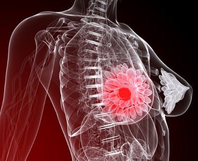
Mastitis is a disease characterized by inflammation of the mammary gland due to the penetration of staphylococci or other pathogenic bacteria through microcracks and wounds. It most often affects breastfeeding women, but the disease can also occur in non-lactating and nulliparous women. This disease is accompanied by very unpleasant symptoms, in later stages there may be discharge of pus. Mastitis may be a consequence of lactostasis.
Causes of the disease:
Symptoms:

Preventive measures
Prevention of mastitis is very important for a woman, because the disease is quite unpleasant and can cause a lot of consequences, especially if the disease has progressed to a purulent stage. In advanced stages of the disease, surgery and pharmaceutical medications may be necessary. If the disease has not yet occurred, it is worth asking yourself how to avoid mastitis.
Prevention of lactational mastitis includes compliance with the following recommendations:

You must always remember that the more natural the process of feeding a child is, the less likely it is to get mastitis. Therefore, there are a number of additional recommendations:

Prevention of non-lactation type of disease
Non-lactation mastitis most often occurs during periods of hormonal disorders, for example, during menopause or menopause in women. Also, disorders may be associated with weakened immunity during puberty or as a result of injuries, operations, or chronic diseases.
Non-lactation mastitis is much easier than lactation mastitis. Therefore, it is necessary for prevention.
Prevention of mastitis in postpartum period, especially during breastfeeding, is of great importance not only for the woman, but also for her child. Mastitis (inflammation of the mammary glands) is one of the infectious diseases accompanied by painful sensations and purulent discharge, which contributes to lactation disorders.
Distinction and classification
There are several types of mastitis. It is classified by paying attention to the nature of the inflammation. Purulent, infiltrative, serous, gangrenous and abscess mastitis are known.
Serous mastitis occurs first. If no measures are taken, it turns into infiltrative, and then into a more complicated form - purulent.
If the disease is not cured for a long time, in the absence of immunity, a complication may occur - gangrenous mastitis.
Once this diagnosis is established, you will have to finish breast-feeding and transfer the child to artificial nutrition. That is why the prevention of mastitis plays an important role for all mothers, so as not to deprive the baby of breast milk, which is necessary for the full development of the baby. But how can you stop breastfeeding without mastitis and what should you do about it? First you need an examination.
Diagnosis of occurrence
The presence of the disease can be determined by examining and palpating the breast. The woman feels pain from the compacted formations.
A more accurate diagnosis is established when ultrasound examination, where inflammatory processes are well observed.
Sometimes a mammogram is performed to clarify the diagnosis. To investigate the presence of infection, milk is taken from a nursing mother from a sore breast.
Prenatal pathology
 In women who are expecting their first child, mastitis occurs quite often. It makes itself felt during the period when the mammary glands begin to swell before childbirth, as well as during the pathological course of pregnancy.
The cause of any mastitis is considered to be an infection - staphylococcus, streptococcus or E. coli. It can penetrate into the breast tissue and contribute to the development of inflammation. The infection can get through cracks in the nipples, scratches and abrasions, and through the lymph. Typically, the development of the disease is associated with decreased immunity.
Another cause of prenatal mastitis is considered to be a hormonal imbalance in the female body. Against this background, it begins to develop fibrous mastopathy. It occurs when a woman has too much of the female sex hormone estrogen and not enough progesterone. This disease can occur in young girls and women early stages pregnancy.
When it occurs, tissue compaction is detected in the mammary glands, which is accompanied by pain. Some of them begin to turn into knots.
Note! If it is detected, you cannot delay the disease; you must visit a specialist - a mammologist. The doctor will prescribe a comprehensive treatment.
Prevention
 You can avoid mastitis during pregnancy by taking precautions. And to prevent mastitis from appearing, prevention is needed, which consists of the following measures:
- wearing a comfortable bra that does not compress the breasts;
- daily washing of the mammary glands with water at room temperature;
- drying with a rough towel;
- taking air baths while lying on the bed with your chest open for 15 minutes a day;
- Breast irradiation ultraviolet rays, to destroy germs (about 15 sessions);
- the need for nipple preparation: massaging, stretching;
- avoiding the appearance of wounds on the mammary glands;
- wearing only clean and ironed underwear;
- taking hormone tests as directed by a doctor.
When a woman comes to the antenatal clinic, conversations should be held on preparing the mammary glands for breastfeeding and how to avoid mastitis.
Postpartum mastitis
 The cause of mastitis in the postpartum period is considered to be stagnation of milk in the glands during breastfeeding, which can occur due to various reasons. These include underdeveloped mammary ducts and impaired nipple formation. Pus begins to accumulate in the milk ducts, the contents of which affect the severity of the disease.
Any type of mastitis, including the occurrence after childbirth, has many common symptoms, namely:
- temperature increase;
- painful sensations in the chest;
- weakness;
- increase in breast size;
- redness of the inflamed area;
- insomnia;
- decreased appetite;
- enlarged lymph nodes under the armpits;
- increased ESR and increased lymphocytes;
- headache.
The disease is diagnosed based on a general blood test, complaints from a nursing mother, the results of an ultrasound examination, and analysis of milk and purulent discharge to identify bacteria.
Prevention
 To avoid mastitis in the postpartum period, you must follow the following rules:
- Before feeding, wipe your breasts with a warm towel or take a shower to improve milk flow;
- learn to feed the baby correctly, that is, make sure that he takes the nipple well and grasps the area around it;
- when feeding, you need to lightly massage the breast, direct movements towards the nipple;
- to soften the nipples, you can lubricate them with vegetable or butter;
- For each feeding you need to give a different breast;
- do not overcool the mammary glands;
- wear the right bra size;
- After feeding, it is necessary to express the rest of the milk completely.
Taking care of your breasts, maintaining hygiene, protecting yourself from accidental blows - all this will help prevent mastitis.
Lactation mastitis and cracked nipples
 This mastitis occurs when there are sources of infection in a woman’s body. It leads to the fact that in a woman’s milk ducts, especially where they are dilated, milk coagulates and stagnates, and the walls swell. Infection in the glands leads to mastitis.
There are acute and chronic mastitis. It is usually initially defined as serous, then develops into more complicated types with purulent accumulations in the milk ducts.
The disease usually occurs a month after birth, and its active manifestation is not observed. This may be characterized by an infiltrative or purulent appearance.
It is mainly diagnosed when body temperature rises to 38°C or higher, when the skin becomes red and the mammary gland increases in volume.
Prevention lactation mastitis First of all, it consists in timely prevention and relief of lactostasis (stagnation of milk in the ducts). When observing lactostasis, you can alleviate the condition of a nursing mother by expressing milk. The pain stops and the temperature returns to normal.
Lactostasis must be stopped within 3 days, otherwise it will be too late. But when measures are not taken in time, mastitis begins to develop. The lump is not removed when expressing milk, and the body temperature rises.
A common cause of any mastitis is the appearance of cracks in the nipples, through which infection easily penetrates.
The causes of cracked nipples are:
- failure to comply with hygiene rules;
- monotonous diet with insufficient amounts of vitamins;
- inept breastfeeding and expressing milk;
- insufficient control of medical personnel in the maternity hospital at the time of breastfeeding the baby.
It is necessary to wash the mammary glands daily with baby soap and wear only clean underwear, and be sure to wear a bra so that the mammary glands are slightly raised to avoid stagnation of milk.
How to stop lactation correctly
 When a child approaches 12 months, his diet, in addition to milk, includes: different types complementary feeding, during this period it is necessary to gradually stop breastfeeding.
Sometimes children, having eaten well with porridge, vegetable purees, and cottage cheese, stop suckling on their own. Then feeding is painless and easy to finish, and the milk becomes smaller each time. But this rarely happens, mostly you have to find different ways, how to stop feeding correctly so as not to harm the baby and mother.
Many mothers don't know when to stop breastfeeding. Some do not feed after 6 months, others do not stop feeding until 2 years. More correctly, this is feeding up to 1.5 years. It is by this time that the milk gradually becomes less. The baby nurses mostly at night or when he falls asleep. Therefore, finding ways to stop feeding will be a necessity.
You should not stop breastfeeding under the following circumstances:
- when a child is teething;
- in summer;
- when is vaccination scheduled?
- during illness;
- if there is a lot of milk;
- when moving to a new place.
But over time, much less milk will still be produced, and then you can think about ending feeding.
Ways to complete feeding
 In order to reduce milk production, you need to buy a bra one size smaller. There is no need to feed your baby at night. The glands should not be heated in a warm shower. Before feeding, it is better to express the milk, leaving a little. Hold the baby less so that he does not feel skin contact.
You can use herbal decoctions that reduce lactation. These include elecampane, lingonberry leaves, sage, horsetail, parsley, mint, and bearberry. All of them have a diuretic effect, and therefore delay the formation of milk. During the day you can drink up to 1.5 liters of herbal decoction.
If you cannot complete lactation without pills folk remedies, medications can help with this.
Important! Close communication with a specialist allows not only to provide adequate treatment. In addition, the doctor will tell you how to finish breastfeeding correctly so that there are no complications.
When taking appointments medicines, the exact dosage must be observed. Such drugs may be Bromocretin, Dostinex. The mammary glands should not be ligated to avoid lactostasis. Having decided to stop breastfeeding your baby, it is important to take this step immediately and no longer put your baby to the breast.
conclusions
Prevention of mastitis allows you to avoid its occurrence. You need to familiarize yourself with the symptoms and treatment methods in advance. It is advisable to prepare the glands during pregnancy for future feeding of the child after birth. And the main thing to remember is that no product can replace the unique mother’s milk.
In the video presented in this article you will find additional information on this topic.
It is important to know! In women who have not given birth under 25-30 years of age, fibrocystic disease (mastopathy) does not cause much concern, but closer to 30, especially during pregnancy and after childbirth, 80 percent of women develop a complication of mastopathy. Along with women who have not given birth, many mothers who devote almost all their time to their baby forget about their health or think that this problem is trivial and will go away on its own. Expectant mothers are in an even more difficult position - during pregnancy and breastfeeding, many pharmaceutical drugs prohibited. Did you know that mastopathy, if not treated in time by preventing the disease, can cause breast cancer. Read about a completely natural remedy for mastopathy (fibrocystic disease), compatible with breastfeeding and pregnancy here...
16. The system for the prevention of mastitis in cows consists of a complex of zootechnical, agrotechnical, veterinary, sanitary and economic measures.
Strict compliance with zootechnical, hygienic, veterinary and sanitary requirements is the main condition for reliable prevention of mastitis in cows.
The basis of general zootechnical measures for the prevention of mastitis is compliance with the rules of milking, zoohygienic standards for keeping and caring for cows, and sufficient and complete feeding of them. One-sided (highly concentrated or silage-pulp) feeding of cows, or feeding them spoiled, moldy, frozen feed, which can cause diseases of the gastrointestinal tract and contribute to the occurrence of mastitis, should not be allowed.
To prevent mastitis caused by diseases of the gastrointestinal tract, at the beginning of the grazing period,
It is recommended to feed cows 1-2 kg of hay or straw at night.
Active exercise is an important preventive measure not only for metabolic disorders, but also for mastitis in cows. Therefore, during the stall period, daily walks are organized for cows over a distance of at least 4-5 km.
Before and after calving, succulent feed is excluded from the cows’ diet and the supply of concentrates is reduced to 1-1.5 kg. It is better to feed cows with good hay at this time. From the 4-5th day after calving, succulent feed is introduced into the diet and by the 10-12th day the feeding level is brought to full normal.
In premises where lactating and dry animals are kept, it is necessary to maintain the required microclimate and sanitary order. Particular attention should be paid to the quality of litter and timely removal of manure. The litter should be moisture-absorbing, warm and soft. Dry straw and sawdust should be used as bedding material.
Ventilation in the barn should provide an air exchange of at least 70-85 m 3 / hour per cow.
To maintain sanitary order on farms, it is recommended to hold a sanitary day once a month. Twice a year in spring and autumn, preventive disinfection of barns should be done. The maternity ward undergoes thorough mechanical cleaning and disinfection every ten days. The passages in the maternity rooms are regularly sprinkled with powdered lime.
Cows are transferred to the maternity ward 10-15 days before calving and returned to the barn 10-14 days after calving. Before transferring cows to the maternity ward, they are cleaned, contaminated areas of skin are washed, and the external genitalia are disinfected with a solution of potassium permanganate 1:1000. When udder edema appears, 2-3 weeks before calving, the cows are limited in the provision of succulent feed and water, and long walks are prescribed.
Proper milking is the most important measure for the prevention of mastitis in cows.
Regardless of the milking method, a thorough pre-milking treatment of the udder is carried out. One minute before putting the teat cups on the nipples, the udder is washed with warm water (temperature 40-45°) from a spray bottle and wiped with a clean towel, and then the lower one is wiped with a clean napkin moistened in a disinfectant solution (0.5% solution of desmol, iodine monochloride). part of the udder and teats. It is advisable to change the napkin after each cow.
In the absence of special devices, it is allowed to wash the udder from a bucket with one of the disinfectant solutions (0.5% chloramine, 0.5% iodine monochloride, sodium hypochlorite or dezmol).
The first streams of milk are poured into a special mug. Coll
It is absolutely unacceptable to milk the first streams of milk on the floor, since secretions from sick cows can cause the spread of infection.
In the prevention of mastitis in cows, the hygiene of milkmaids is of great importance. Before milking, the milkmaid puts on a clean robe, washes her hands with soap and wipes them with a clean towel.
All farm workers involved in milk production must periodically undergo a medical examination and carefully comply with the requirements set out in the “Sanitary and Veterinary Rules for Dairy Farms of Collective and State Farms for the Care of Milking Plants, Equipment and Dairy Utensils and Determination of the Sanitary Quality of Milk”, approved by the Main Veterinary Directorate of the USSR Ministry of Agriculture on January 22, 1970.
In order to prevent mastitis during machine milking, special measures should be taken, which include:
a) preparing farms for machine milking;
b) monitoring the condition of the udder of cows;
c) monitoring the correct operation of milking machines;
d) control over the sanitary condition of dairy equipment.
PREPARING FARMS FOR MACHINE MILKING
17. Milkmaids, cattlemen, mechanics and other farm workers must undergo a theoretical course and practical training in the techniques of machine milking of cows and sanitary processing of milking equipment.
Healthy cows whose udder shape meets the relevant requirements are transferred to machine milking. The most desirable shape of the udder is bath-shaped, cup-shaped with medium-sized (5-9 cm) cylindrical teats located at right angles to the udder.
Herds in which about half of the cows have an udder shape that does not meet the requirements of machine milking, it is better to transfer to less dangerous milking with Volga three-stroke milking machines.
Herds in which two-thirds of the cows have an udder shape suitable for machine milking can be transferred to milking with two-stroke milking machines DA-2 “Maiga”, “Impulse”.
Farms with machine milking of cows must have equipped dairy, cold and hot water, detergents and disinfectants, as well as an uninterrupted supply of electricity.
Installation of the milking machine must be carried out in accordance with the technical specifications. The assembled milking machine is accepted by a commission consisting of representatives of the farm, V/O Soyuzselkhoztekhnika, the chief specialist in livestock mechanization of the district agricultural department, as well as the district veterinary service.
When accepting the installation of a milking machine, special attention should be paid to the compliance of the operating modes of the milking machine and individual milking machines in accordance with the requirements of the operating instructions.
CONTROL OF THE CONDITION OF THE COW'S UDDER
18. When machine milking cows, monitoring the condition of the udder is decisive in the prevention of mastitis.
When milking the first portions of milk onto the black strainer of the mug, the milkmaid pays attention to the color of the milk, the presence of flakes, blood clots, mucus and other inclusions in it.
During milking, the milkmaid must monitor the behavior of the cow. The animal's restlessness, stepping from foot to foot, an attempt to throw off the milking machine, and milk retention indicate an irritating effect of the machine or an udder disease.
After milk production stops, the apparatus is immediately removed so as not to overexpose the glasses to the milked udder. The milkmaid turns off the vacuum and only after that removes the glasses from the teats by lightly tugging on the manifold. You cannot remove the teat cups without turning off the vacuum, as this leads to ruptures of the mucous membrane and disruption of the integrity of the teat skin.
After finishing milking, the milkmaid must examine the cow's nipples and udder. Any deviations from the norm are immediately reported to the foreman, veterinarian or livestock specialist.
As planned, all dairy cows are regularly checked for hidden mastitis at least once a month using a 5% solution of dimastin or 2% mastidine.
Cows suffering from latent mastitis are milked by hand last, taking precautions to prevent the spread of infection to other cows, and milk from sick animals is not allowed to be mixed with general milk.
Boiled milk from cows with mastitis is used only for animal feed. If there is a significant change in the milk (presence of pus, fibrin, blood), it is destroyed.
If mastitis is widespread in cows, after milking it is necessary to immerse the tips of the teats in a 0.5% solution of iodine monochloride, desmol, or lubricate them with an antiseptic emulsion. A glass is filled with a disinfectant solution, into which the ends of the nipples are placed without subsequently wiping them.
CONTROL OF THE OPERATION OF MILKING MACHINES
19. Before starting milking, the milkmaid must check each milking machine for pulsation frequency. In three-stroke devices, the number of pulses should be 60 per minute, in two-stroke devices - 80 ± 5. More frequent pulsation may cause mastitis in cows.
The vacuum value in the vacuum line during milking should be in the range of 380-400 mm for three-stroke machines and 360-380 mm Hg for two-stroke machines. The vacuum value in the milk pipeline of the milking plant milk pipeline-100 (200) “Daugava” is 450-500 mm Hg.
The milkmaid must constantly monitor the position of the teat cups during milking. If the glasses are detected creeping onto the udder, they are pulled forward and down by the collector. A strong creep of teat cups onto the udder is observed in three-stroke machines converted to work using the push-pull method.
Milking with such machines causes a large number of cows to develop mastitis, so converting three-stroke machines to a two-stroke mode of operation is unacceptable.
When milking into the milk line, sometimes there is a strong fluctuation in the vacuum in the teat space, which leads to slow milking, collapse of teat cups and reverse flow of milk into the teat cups, causing so-called “wet milking”. This contributes to the transfer of infection from sick to healthy lobes of the udder.
If the teat cups fall off during milking, you should pay special attention to the vacuum mode of the installation or to the condition of the vacuum wire, which may be clogged.
When the milking cups fall off, you must immediately turn off the milking machine, wash them thoroughly in a hot 0.5% solution of sodium hypochlorite, desmol, rinse with hot water and only then continue milking.
During the milking process, it is necessary to ensure that the cows are completely milked by the machine. As soon as the flow of milk has stopped, it is necessary to switch to machine milking. For this purpose, the milking cups are pulled forward by the collector for 25-30 seconds. If during this time the milk flow has not increased, then stop milking and remove the glasses.
When milking cows with an udder shape that does not meet the requirements of machine milking, hand milking is allowed in some cases (different times for milking quarters).
A very important preventive measure is to determine the end of milk production in order to avoid dry milking, which leads to mastitis in cows. If no more than 250 ml of milk remains in the udder after milking, then the cow has been milked well. With an increase in the amount of unmilked milk, self-starting of cows and an increase in mastitis are observed.
CONTROL OF THE SANITARY CONDITION OF DAIRY PRODUCTSEQUIPMENT
20. To prevent mastitis, it is necessary to strictly observe sanitary regimes for the care of milking machines and milk utensils.
Veterinary specialists must carry out systematic monitoring of the sanitary condition of milking installations, paying attention to contamination of teat rubber, collector, milk hose, lids and gaskets of milking buckets, viewing devices, milk pipes, filters, coolers and milk pumps, which are most contaminated during operation . When parts of the milking machine become dirty, they become covered with grayish mucus or even milk clots.
SANITARY TREATMENT OF MILKING MACHINESAND DAIRY DISHES
21. The following modes are recommended for sanitizing milking machines with a milk line, portable machines and milk utensils.
MODE I. After each milking, rinse with warm water to remove any remaining milk, then wash with a hot 1% sodium hypochlorite solution and rinse again with warm water.
MODE II. After each milking, rinse with warm water, wash with a 0.5% solution of washing powder A, B or C and disinfect once a day with a 0.1% solution of sodium or calcium hypochlorite or chlorine water. After disinfection, rinse with warm water.
MODE III. After each milking, rinse with warm water, treat with a 0.5% solution of desmol and rinse with warm water.
MODE IV. Rinse with warm water, treat with a hot 0.5% solution of powder A, B, C or soda ash. Once a day, disinfect with steam for 3 minutes.
MODE V. For sanitary treatment of milking equipment using a circulating washing stand, use 0.25% solutions of detergents and 0.1% solutions of disinfectants in the above sequence.
In each farm, based on specific conditions, a specialist can choose the most appropriate mode of sanitization of milking equipment, which are recommended by these rules.
An indicator of the effectiveness of sanitary measures carried out on farms is also the results of milk testing for mechanical and bacterial contamination, which is carried out every ten days by dairy laboratories.
PREVENTION OF MASTITES DURING THE DRY PERIOD
22. Cows are started l"/g-2 months before the expected calving. At the same time, the supply of succulent feed and concentrates is limited to 50% of the diet. From three times milking they switch to double milking, then to single milking, after which they milk every other day and stop milking. Cows that are difficult to start are, in some cases, completely succulent and concentrated feeds are excluded from the diet and watering is limited.
The disease of cows with mastitis with a subclinical course is observed quite often during the start-up and dry periods. If there is no control over the condition of the udder during this period, mastitis goes unnoticed and is discovered after calving. In many cases, postpartum mastitis is a consequence of infection of the udder during dry periods. Therefore, during the dry period, it is recommended to conduct a clinical examination of the udder with a test milking of secretions once every two weeks. In order to prevent mass postpartum mastitis, cows suffering from subclinical mastitis can be intrauterinely administered long-acting antibiotics (bicillin-3) at a dose of 300 thousand units in each quarter of the udder or preparations mastikur, masticide, according to current instructions. In this case, antibiotics are administered after testing cows for mastitis through a test milking, test with dimastin and settling.
Antibiotic therapy for subclinical mastitis during the dry period reduces the number of clinically significant postpartum mastitis in cows by 50%.
|
ANNEX 1 |
The secret of the sick quarter |
Initially unchanged in appearance, and then liquid, often with flakes |
The secretion is liquid, watery, grayish-white in color, mixed with yellowish or whitish flakes. Less commonly, a small amount of yellowish whey with flakes or a thick, creamy mass is released |
Serum with fibrin crumbs; less often, the exudate is thick with a predominance of flakes. There may be blood impurities, blood clots, and tissue fragments |
|
Differential diagnosis of acute mastitis in cows |
Quarter condition |
More often than not, half or all of the udder is affected. It is enlarged, unevenly compacted, and doughy in places. The nipple is often enlarged and swollen |
One quarter is affected, it is enlarged or compacted in areas, especially at the base. The nipple is unchanged, less often edematous, with foci of fluctuation at the base |
One quarter is affected. It is sharply enlarged, compacted with the presence of individual dense nodes and areas of softening. The nipple is swollen. Sometimes crepitus is noted |
|
Skin condition local temperature, udder soreness |
The skin is tense, non-pigmented areas are hyperemic, the temperature is elevated, the pain is significant |
The skin is unchanged, the temperature is rarely slightly increased, the pain is mild or not at all |
The skin is tense, non-pigmented areas are hyperemic. Body temperature is increased. A quarter is painful |
|
|
General condition of the animal |
Often no change, less often oppression; body temperature is normal or slightly elevated, sometimes lameness |
No visible changes, sometimes slight depression; decreased appetite; increase in body temperature |
Depression, decreased or lack of appetite; increased body temperature; lameness, emaciation |
|
|
Serous mastitis |
Catarrhal mastitis |
Fibrinous mastitis |
|
The secret of the sick quarter |
The exudate is mucopurulent, often thick with white or yellow flakes, can be liquid, yellow-red in color |
At first, the milk does not change in appearance, but there is little of it. Then it becomes watery, grayish-white or yellowish in color, and contains an admixture of pus and casein. The type of secretion depends on the number of abscesses and the location of their opening (through the skin or into the lumen of the milk ducts) |
There is little secretion, it is watery, grayish in color with an admixture of flakes, and often bloody |
There is little secretion, it is watery, reddish in color, with flakes and blood clots |
|
Quarter condition |
The affected quarter of the udder is enlarged and in some places has compacted lesions. The nipple is sometimes swollen |
The affected quarter is unevenly enlarged, fluctuating, tense foci of varying sizes are palpable. With deep location of abscesses, the quarter is enlarged, fluctuation is weakly expressed |
The affected quarter is significantly enlarged; very swollen nipple |
More often, half or all of the udder is affected. The affected part is uniformly enlarged and compacted. Swollen nipple |
|
Skin condition, local temperature, udder soreness |
The skin is tense, non-pigmented areas are hyperemic. body temperature is increased. Soreness is expressed |
The skin at the site of the outbreak is tense, swollen, hyperemic, hot, painful |
The skin is tense, swollen, uniformly hyperemic, temperature and pain are significant |
The skin is swollen evenly, diffusely hyperemic, the temperature is elevated, the pain is significant |
|
General condition of the animal |
Depression, refusal to feed; increased body temperature; lameness |
Depression, loss of appetite, significant increase in body temperature; with multiple abscesses, remitting fever; lameness |
Severe depression, significant increase in body temperature; decreased or lack of appetite; lameness |
Depression, significant increase in body temperature; decreased appetite |
|
Purulent catarrhal mastitis |
Udder abscess |
Phlegmon |
Hemorrhagic mastitis |
APPENDIX 2
LABORATORY RESEARCH METHODS
 Serous mastitis is a postpartum inflammation of the mammary gland in women, occurring with a frequency of up to 20% of the total number of births. (according to Professor L.N. Bisenkov and staff of the Department of Thoracic Surgery of the S.M. Kirov Military Medical Academy (St. Petersburg, 2014). Without appropriate treatment, this disease can be complicated by the formation of ulcers in the tissue mammary gland which can only be treated surgically. At the same time, there are risks of depriving the baby of breast milk and other irreversible consequences.
What is postpartum mastitis, what are the causes, symptoms and diagnosis of serous (lactation) mastitis, how is inflammation of the mammary glands treated in women, does serous mastitis affect the child and is it possible to continue breastfeeding - you will read the answers to these and many other questions in this article.
In itself, lactostasis is not a disease. But it must be eliminated for further successful breastfeeding. Most The best way To do this, do not stop feeding, because a hungry baby, while sucking, will empty the breast and use up the entire supply of milk. Pumping, massage, and physiotherapy are also very effective. Physiotherapy for lactostasis and its prevention are carried out.
You can read a detailed story about lactostasis, methods of its elimination and prevention, including a detailed description of pumping and massage techniques.
But, nevertheless, lactostasis remains only lactostasis and is not a disease until infection of the stagnant milk and adjacent breast tissue occurs. From this point on, we can talk about inflammation of the mammary gland in women - lactation mastitis.
During mastitis, three stages are distinguished - serous (serous mastitis), infiltrative and purulent.
Each stage is regarded as an independent form of lactation mastitis.
If inflammation of the mammary gland in a nursing mother is at the serous stage of development, then a diagnosis of “Acute serous mastitis” will be made.
The infiltrative and purulent stages of the disease in the medical history will be designated as “Acute infiltrative mastitis” and “Acute destructive mastitis.” In the latter case, the name comes from the word “destruction” - destruction, because During the purulent stage, melting of the tissue structures of the mammary gland occurs.
Mechanisms of development of lactation mastitis
Serous stage or serous mastitis.
 After infection, pathogens located in the tissues of the gland begin to multiply, especially since stagnant milk is an excellent nutrient medium for this. At the same time, they release their waste products, which are toxic to the body. The first symptoms appear - malaise, weakness.
In response to the introduction of infectious agents, it is activated the immune system and its effector cells, leukocytes, whose task is to destroy pathogens, enter and accumulate at the site of inflammation. All this is accompanied by dilation of microvessels, the transition of the liquid part of the blood into the tissue and the formation of edema.
Stagnant milk, accumulation of microorganisms and leukocytes, dilated microvessels, edematous fluid - all this together forms a focus of inflammation, the local signs of which are swelling (hardening), local increase in temperature, redness, pain, dysfunction. Local manifestations are accompanied by general ones - increasing malaise and weakness, fever, changes in the cellular composition of the blood (reflected in general analysis blood).
If treatment is insufficient or absent altogether, the serous stage can transform into infiltrative and purulent.
Infiltrative stage or acute infiltrative mastitis.
 As the disease progresses, the composition of the inflammatory fluid changes. First this serous the liquid is clear with a small amount of leukocytes and protein. Then many dead leukocytes, decay products of tissues and protein molecules appear in it. Now it is a viscous yellow liquid - pus, which is actively spreading, being implemented into neighboring, previously undamaged structures.
The process of spread is called infiltration, hence the name of the stage - infiltrative, as well as the form of the disease - acute infiltrative mastitis.
A contributing factor to infiltration is the structural features of the mammary gland, in the tissues of which there are no dense connective tissue partitions between its individual parts. Therefore, the spread of purulent inflammation of the mammary gland occurs quite quickly.
Infiltration into previously healthy tissues leads to the formation of accumulations of pus. From now on they talk about the purulent stage postpartum mastitis– acute destructive mastitis.
Purulent stage or acute destructive mastitis.
 Infiltration into previously healthy tissues leads to the formation of accumulations of pus, which can be in the form of abscesses or phlegmons.
An abscess is an abscess enclosed in a capsule. There may be different locations of abscesses (purulent inflammation of tissues) - in the peripapillary zone, under the skin, in the thickness of the mammary gland (intramammary form), behind the gland (retromammary form). If the abscess is not delimited by a capsule from healthy tissue, then it is phlegmon.
The purulent stage or acute destructive mastitis is much more severe than the serous and infiltrative form and requires surgical removal accumulations of pus, is dangerous for the spread of infection beyond the affected breast, up to the development of blood poisoning.
Symptoms and signs of serous (lactation) mastitis
Lactation mastitis is always preceded by. IN mammary gland a lump appears that can be painful. Feeding a baby can also be accompanied by pain. Milk is released slowly, in drops, with difficulty. The skin over the lump turns red. Often there is a general malaise, an increase in body temperature of 37.4-37.6 ° C, and the temperature will be higher in the armpit, which is located next to the affected gland. After pumping or completely emptying the breast, the child’s general condition improves significantly and the temperature also drops.
If an infection is added to the stagnation of milk, then lactostasis turns into serous mastitis. The general condition worsens, weakness, headaches, chills, and often nausea and vomiting appear. The temperature rises to 38.0-39.5 ° C, does not normalize after pumping and fluctuates repeatedly during the day with a difference between the maximum and minimum values of more than 1-2 ° C - debilitating fever. When measuring temperature, there is no difference in the right and left armpits.
If left untreated, serous mastitis progresses to infiltrative and then purulent form. There is a progressive deterioration in the general condition with a rise in temperature to 39.0-40.0 ° C, increasing induration, engorgement and swelling of the mammary gland, severe pain in the area of induration. Changes in the general blood test are increasing.
Diagnostics
 When the first signs of inflammation of the mammary gland appear in a nursing mother, you need to consult a surgeon who will make a diagnosis and, at the same time, exclude other diseases similar to mastitis.
At the appointment, the doctor will listen to your complaints, ask a number of clarifying questions, perform a general examination and a special local examination of the affected gland.
Then he will order an examination, which may include:
- General blood test - to identify nonspecific signs of an inflammatory reaction, which are expressed in an increase in the total number of leukocytes more than 9.0x10 9 /l (leukocytosis), the relative content of neutrophils over 75% (neutrophilia), erythrocyte sedimentation rate more than 20-30 mm/hour ( increase in ESR).
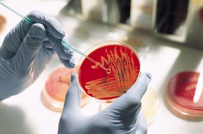
- Bacteriological research milk - is carried out by inoculating this liquid on nutrient media. If microorganisms are present in milk, they form their own colonies on the media, based on the characteristics of which the type of pathogen can be determined. At the same time, its sensitivity to antibiotics is determined and this study is performed within 5 days. Therefore, until the result is obtained, patients are prescribed broad-spectrum antibiotics. Below, we will talk in more detail about antibiotic therapy in the treatment of serous mastitis.
- Ultrasound diagnostics(Ultrasound) is prescribed to distinguish between mastitis and similar diseases - cystic mastopathy and breast cancer.
- Computed mammography is also used to differentiate serous lactation mastitis from cystic mastopathy and breast tumor.
- Breast thermography (thermomammography) – records infrared radiation from areas of the body. If lactation mastitis is suspected, it is used to exclude breast cancer, which is accompanied by much more intense radiation.
Treatment of inflammation of the mammary glands in women
When planning treatment measures, the doctor is primarily guided by the following mandatory rule: acute serous and infiltrative mastitis is treated conservatively (i.e. without surgical intervention), and purulent and purulent-destructive forms - surgically.
Effective conservative treatment of serous mastitis must be timely, rational and comprehensive.
The complex of treatment measures includes bed rest, frequent pumping, continued breastfeeding and mandatory physiotherapeutic procedures.
- Bed rest. This means that a woman with lactation serous mastitis is allowed to sit up in bed and turn around, but toileting and eating is possible only with the help of caregivers. We draw your attention to the fact that independently visiting the toilet, walking around the room (ward), eating at the table is ward rest, but not bed rest, which is prescribed. This increased attention to the regimen is due to the fact that serous mastitis is an active infectious process that can be complicated by cardiovascular disorders. Bed rest limits stress and prevents such disorders.
- Wearing underwear that does not compress the chest because Compression of the breast tissue by a tight bra interferes with the outflow of milk from it.
- Frequently expressing milk from the affected breast– a mandatory requirement, because, firstly, stagnant milk curdles and begins to ferment, and this is an excellent breeding ground for pathogens of postpartum mastitis; secondly, accumulations of milk compress the neighboring milk ducts, blood vessels and gland tissue. To eliminate milk stagnation, it is very effective (see below), which allows you to quickly express milk and eliminate disturbances in the outflow tract (from (St. Petersburg, 1997)).
- Lactation in conditions of serous inflammation of the mammary gland continues. Serous mastitis is not a contraindication for this. On the contrary, when the baby sucks, he quickly eliminates milk retention and completely empties the breast.
- Reducing the intensity of milk production to “unload” congestion. For this purpose, it is recommended to slightly reduce the amount of liquid you drink per day.
- - aimed at accelerating the cleansing of tissue from damaged cells, decay products and vital activity; improving cell nutrition, better penetration of immune cells, their contacts with infection. By accelerating all these processes, swelling and inflammation are relieved more quickly. "Vitafon", unlike medications inhibition, blocking and reduction of inflammatory reactions (edema), helps the body quickly solve the problem of recovery and, thus, swelling and inflammation go away on its own, but only faster than usual. As a result, it is the most natural way for the body to heal.
The Vitafon device can also be used at home. In this case, the source of inflammation is exposed to microvibrations - modulated mechanical vibrations of low intensity in the acoustic frequency range. What explains the therapeutic effect of vibroacoustic therapy?

Thus, in case of serous mastitis, vibroacoustic therapy with the Vitafon apparatus effectively eliminates milk stagnation and swelling, normalizes blood supply to the mammary gland, and enhances the destruction of pathogenic microorganisms.
We bring to your attention doctor's video review highest category, candidate of medical sciences, associate professor of the department of pediatrics of Northwestern State Medical University named after. Mechnikova (St. Petersburg) F.N. Ryabchuk on the use of the Vitafon device for lactostasis and serous mastitis.
9. Antibacterial drugs - are prescribed in accordance with the National Guidelines for Clinical Surgery (2008) (see list of references) in the form of a course of intramuscular injections. Before the beginning antibacterial therapy The sensitivity of the pathogen to the drugs must be determined. The study takes 5 days and during this period of time the patient receives broad-spectrum antibiotics. After receiving the result, a drug is prescribed that selectively destroys the type of pathogen (strain) that was identified.
In this case, the following questions often arise: Is it possible to continue breastfeeding while taking antibiotics? Will taking antibiotics harm my baby?
Answer: yes, it is possible to breastfeed a child while taking antibiotics in conditions of serous mastitis, and taking antibiotics will not harm the child if the following circumstances are taken into account when prescribing antibacterial therapy for serous mastitis:
- firstly, maintaining breastfeeding even in conditions of serous mastitis remains a paramount task;
- secondly, for treatment with antibiotics and simultaneous breastfeeding, drugs should be selected that do not penetrate into milk or are present there in trace concentrations;
- thirdly, the chosen drug must be absolutely non-toxic (this applies to those antibiotics that are found in milk in trace quantities);
In accordance with the criteria specified here, all antibiotics are divided into permitted (only they can be used), conditionally permitted, prohibited and unstudied:
- Allowed, which can really be taken with caution by nursing mothers with mastitis. These include a small number of drugs from the penicillin group. Never try to decide on your own which specific antibiotic to choose. This issue is entirely within the competence of the doctor.
- Conditionally permitted antibacterial drugs– a group of aminoglycosides - are prescribed when they cannot be avoided, i.e. benefit outweighs harm. These are the cases when a woman breastfeeding her child develops diseases such as meningitis, sepsis, etc.
- Strictly prohibited antibiotics - tetracycline, chloramphenicol, ciprofloxacin, lincomycin, metronidazole, clindamycin, fluoroquinolone antibiotics.
- Unexplored There is no data yet on their effect on the child’s body. They should not be taken while breastfeeding.
Prevention of lactation mastitis in the postpartum period
We present to your attention a video from useful tips for the prevention of mastitis.
Measures to prevent the development of the inflammatory process in the mammary glands are planned and carried out in the prenatal period, during and after childbirth.
During the prenatal period eliminate pregnancy complications that can reduce resistance female body women to the action of pathogens. These complications are identified during routine visits to the gynecologist. The doctor will also take care of the timely sanitation of chronic foci of infection - carious teeth, chronically inflamed tonsils, old inflammations in the urinary tract, etc.
Special preparation of the breasts and nipples for breastfeeding is needed only when the nipples are flat and the areola is tight and not stretchable.
 To determine the shape of the nipple, place it between your thumb and forefinger and squeeze lightly. The convex nipple will look forward, and the flat or inverted nipple will “hide” between the fingers. In such cases, nipple massage is used. You can do this massage yourself at home, but only after your doctor approves this procedure, because exposure to the nipples can cause contractions of the uterine muscles. To massage, you need to grab the nipple between two fingers and gently pull it out, slightly twisting it or rolling it between your fingers. The procedure should last no more than one minute.
Immediately after birth Follow the rules for preventing postpartum mastitis every day:
- To prevent stagnation of milk (lactostasis), feed the baby “on demand” and without time limits, do not allow feeding in the same position, make sure that the baby grasps the nipple correctly, etc. You can read the full description of the necessary actions in the article “”.
- Carry out preventive maintenance periodically. This procedure completely prevents the retention of milk in the outflow tract and increases the activity of immune antimicrobial defense in the breast tissue.
- Immediately after birth, throughout the entire lactation period, after each feeding, apply a thin layer to the nipples and lightly rub in Bepanten cream. It is used during lactation for the prevention and treatment of cracks and redness of the nipples.
- Breast hygiene also affects the appearance of cracks. There is no need to wash, let alone soap, your breasts and nipples before and after each feeding. This washes away the protective layer of oils, dries out the skin and can lead to cracks. It is enough to take a shower 2 times a day.
- Make sure you are properly latched to the breast:
- During feeding, it should be comfortable for both you and the baby.
- The nipple is in the baby's mouth and the lower lip should be slightly turned outward.
- If you need to interrupt feeding (you are in pain, or the baby has not latched onto the breast correctly), then you should not pull out the baby’s nipple, but carefully “screw” your little finger into the baby’s mouth. This creates a space through which the nipple can be removed. While you are doing this, make sure once again how much grip the kids have! If you remove the breast incorrectly, this can also lead to cracked nipples.
- Buy special nipple covers that are worn under a bra - milk will accumulate in the cover, infection will not penetrate the glands, and the nipple will be reliably protected from mechanical damage.
- Use bras one size larger than necessary. They should be pitted and foam-free. This is necessary in order to prevent compression of the chest, irritation and microtrauma of the skin (prevention of cracks).
Conclusion
 The main goal is to maintain breastfeeding and restore lactation at any cost!
Effective treatment of serous mastitis in women must be timely and comprehensive. To do this, physiotherapeutic procedures () must be combined with frequent pumping and not stop breastfeeding.
Preventive measures are carried out throughout the entire period of lactation. This will help prevent stagnation of milk and the penetration of pathogens into the milk ducts of the breast. In addition, they will ensure the destruction of the pathogen if infection does occur.
List of used literature
- Ailamazyan E.K. Obstetrics: textbook. – Moscow State medical University them. THEM. Sechenov. – 9th edition, revised and expanded – Moscow: GOETAR-Media. – 2015
- Obstetrics: national guidelines / Edited by E.K. Ailamazyan, V.I. Kulakova, V.E. Radzinsky, G.M. Savelyeva. – Moscow: GOETAR-Media, 2009
- Bisenkov L.N. Thoracic surgery. Guide for doctors. - St. Petersburg: Hippocrates, 2004.
- Gynecology. National leadership / Edited by V.I. Kulakova, G.M. Savelyeva, I.B. Manukhina. – GEOTAR-Media. – 2009
- Clinical surgery. National leadership in 3 volumes / Edited by A.I. Kirienko, V.S. Savelyeva. – GEOTAR-Media. – 2008 – vol. 2.
- Yakovlev Y. Ya., Manerov F. K. Lactostasis and lactation mastitis in pediatric practice / Siberian Medical Review - 2015 - No. 2 (92) - p. 32-41.
You can ask questions (below) on the topic of the article and we will try to answer them competently!
Mastitis is an inflammation of mammary gland (MG) tissue, regardless of the nature of the process. It can occur due to stasis of milk, nipple cracks, as well as against the background of ectasia of the milk ducts, decreased immunity. As a rule, inflammation affects one or more lobes of the mammary gland. The main symptoms are lumps, pain, local fever, hyperemia, weakness, chills, and discharge from the nipple. There are diffuse and nodular forms of mastitis.
Lactation mastitis is a common pathology of the mammary glands in postpartum women. Their frequency currently amounts to 0.5-5% in relation to the total number of births. Violation of the basic principles of preventing the disease, untimely and improper treatment of its initial forms contribute to the development of severe purulent lesions of the mammary gland, complicated by sepsis. Therefore, the main prerequisite for the correct prevention of mastitis and improving the outcomes of their treatment is maximum attention to this issue on the part of medical workers in obstetric, gynecological and surgical institutions and the consistent implementation of a number of well-founded and practice-tested measures.
Mastitis occurs due to infection of the mammary glands by pathogenic microbes, mainly staphylococci. Predisposing factors to the development of inflammation in the gland are stagnation of milk, the appearance of cracked nipples suffered during pregnancy infectious diseases, complicated course of childbirth, violation of hygienic principles of feeding the child, lack of proper sanitary and hygienic conditions in maternity wards and insufficient compliance with the principles of preventing mastitis at home. Therefore, prevention of mastitis should begin during pregnancy, continue during the woman’s stay in the maternity hospital before childbirth, during the labor and postpartum periods and after discharge from the maternity hospital at home under the supervision of obstetric and gynecological service workers.
Anatomy and physiology of the breast
The mammary glands are located on the anterior chest wall at the level of the II-VI ribs. The parenchyma of the mammary gland is represented by complex alveolar tubular glands, collected in small lobules, from which large lobes are formed. According to various authors, the mammary gland has from 6 to 24 lobes, separated from each other by connective tissue septa. Each of them has its own excretory duct; some ducts may merge before exiting to the surface of the nipple, so the number of holes on the nipple may fluctuate. At the level of the edge of the areola, each duct expands fusiformly, forming the so-called lacteal sinuses. The lining of the glandular structures of the mammary gland is heterogeneous: the alveoli are lined with cubic epithelium, the ducts throughout are cylindrical, and at the mouths (in the nipple) they are lined with multilayered squamous epithelium. The mammary glands are enclosed in connective tissue cases formed by splitting the superficial fascia, and their tissue is penetrated by connective tissue septa extending from the back wall of the case and penetrating into the deep layers of the skin in the form of cords called Cooper's ligaments. Between the fascial capsule of the gland and the fascia of the breast itself there are retromammary fiber and loose connective tissue.
Blood supply to the mammary glands is carried out through the branches of the internal thoracic (about 60%) and axillary (about 30%) arteries, as well as through the branches of the intercostal arteries. The veins of the mammary gland accompany the arteries and widely anastomose with the veins of the surrounding areas.
Throughout a woman’s life, starting from the moment of her birth, the morphological and functional state of the mammary gland changes depending on age, pregnancy and lactation.
The mammary gland of a newborn is 4 times larger than that of a fetus in the ninth month of pregnancy. Sometimes a small amount of colostrum-like fluid is released from the nipple of enlarged mammary glands in newborns. The dilated ducts contain a significant amount of secretion, and their terminal parts give rise to sprouts that are similar to acinci. All these phenomena undergo reverse development within a few weeks or months. The size of the mammary gland in newborns decreases, the expansion of the terminal sections of the milk ducts disappears, discharge from the nipple stops, and the gland becomes calm. This secretory activity of the mammary glands of both sexes is the result of the entry into the child’s body of estrogenic hormones from the mother’s body, which are produced by the placenta during pregnancy. Prolactin, which enters the newborn’s body both immediately before and especially after childbirth, also plays a major role in the occurrence of this phenomenon. Only later, with a decrease in the amount of milk received (after 4-5 months) or a complete cessation of feeding, do the child’s mammary glands become intact.
From the first year of life until the onset of puberty, between 13 and 15 years, the mammary gland, like the ovary, is at rest.
Shortly before the onset of menstruation, and especially after its onset, pronounced changes in growth, development and proliferation of the main tissues of which it consists occur in the mammary gland. A woman's menstrual function begins at the age of 11-15 and continues until the age of 45-52. Repeated at regular intervals of 21-30 days, menstruation is cyclical in nature and externally manifests itself in the form bloody discharge from the genitals.
The development of epithelial and fibrous tissue in the mammary gland, enlargement of the milk ducts, their branches, the appearance of acini and hypertrophy of the epithelium lining the walls of the ducts that occur after the onset of menstruation are the result of the influence of both ovarian hormones and other internal secretion organs. But the ducts and acini of the mammary gland reach their maximum development during feeding, when its function is maximally manifested.
The menstrual cycle in adult women is accompanied by a number of changes in the mammary gland. In the premenstrual period, pain, tension, swelling and even an increase in size often appear in it. For some, this occurs 3-4 days before the start of menstruation, and for others even earlier - from the middle of the cycle. Sometimes there is a small discharge of colostrum from the nipple. After menstruation, all these phenomena disappear.
Thus, cyclical changes in the mammary gland occur mainly in the second half of the menstrual cycle. In this phase, in breast tissue prepared by estrogen under the influence of progesterone and lactogenic (luteotropic) hormone, i.e. prolactin, processes of proliferation and secretion occur with corresponding morphological manifestations. During menstruation, all these processes end and the reverse development of breast tissue begins.
Already 5-6 weeks after the start of pregnancy, an enlargement of the mammary gland is noted. It increases especially quickly from the middle of pregnancy. Superficial veins become very thick and translucent, and striae (white stripes) appear on the skin. The nipple increases in size and its epidermal cover thickens. The diameter of the areola increases and it becomes darker in color. During the first three months (first trimester), there is proliferation of the terminal duct epithelium and formation of sprouts. Wandering cells and young fibrocytes appear. In the second trimester of pregnancy, the terminal ducts sharply increase in number and form large lobules. Their lumens are expanded, acini are formed, lined with cuboidal epithelium, fat droplets are contained inside the epithelial cells, and a small amount of secretion is noticeable in the acini. The connective tissue surrounding them is soft and contains a cluster of lymphocytes. In the third trimester of pregnancy, the newly formed acinci expand, and significant secretion is detected in them. Interlobular connective tissue is rich in blood vessels. There is an increased proliferation of both ducts and acini. Many acinci have low cuboidal epithelium with secretory vacuoles. Some acinci are very dilated, and the beginning of lactation is detected in them.
The mammary gland reaches its most complete development, both functionally and morphologically, during lactation. During lactation, the lobules and their excretory ducts perform a dual function: a) milk secretion and b) milk storage.
Changes after lactation. Regressive changes in the mammary glands occur already at the end of the lactation period. The amount of secretion decreases in the 9-10th month of feeding. These regressive changes reach their most complete expression after cessation of feeding. Acincs subside, the lumen of the ducts decreases. The periductal and interlobular connective tissue regenerates. After lactation, the breast becomes flaccid, since stromal regeneration does not occur fully.
Hypertrophy of the mammary glands depends on age, various disorders of hormonal correlations, which may be caused by diseases of the ovaries and other internal secretion organs.
Significant enlargement of the mammary glands is common in girls and may be part of the precocious ripening syndrome. It has been established that most girls reach puberty by the age of 11-15 years. But there are children whose first signs of maturity appear in the 2nd and 5th years of life. This is expressed primarily by hypertrophy of the mammary gland and the appearance of pubic hair. Menstruation comes later. The cause of premature ripening is often ovarian tumors with pronounced hormonal secretion. Along with this, there may also be constitutional premature maturation without the presence of tumors. A study of hormones in these children showed that they have increased excretion of gonadotropins, estrogens and 17-ketosteroids in the urine. Sometimes the cause of premature development of the mammary glands is tumors of the adrenal cortex and damage to the third ventricle of the brain.
The most common type of true mammary hypertrophy is that which occurs during the period of maturation simultaneously with the appearance of menstruation. In this case, both mammary glands are most often affected, but cases of unilateral damage are also observed. For 1-2 years of strong growth
mammary glands they reach a significant size and, due to their heaviness, fall onto the stomach. Patients experience great discomfort due to the severity of these hypertrophied mammary glands; pain occurs in the shoulder girdle and neck area, and swelling and diaper rash occur under the mammary glands. Microscopic examination of hypertrophied mammary glands reveals only a large increase in the amount of fibrous tissue. Reverse development of this hypertrophy is usually not observed. In some patients, there is a violation of hormonal correlations.
In women who have given birth and breastfeeding a lot, especially in older age, the elasticity of the mammary gland tissue sharply decreases and the ligaments that support it are stretched. At the same time, atrophy of the glandular tissue is observed, and often fat deposition, causing the breasts to sag.
Mammary glands are glandular hormone-dependent organs that are part of the reproductive system women who develop and begin to function under the influence of a whole complex of hormones.
Among estrogens, estradiol plays the most important role in the functioning of the mammary gland. It controls most morphological changes in breast tissue, affecting its tissue, regardless of age. Its concentration in connective tissue mammary gland at 2-20
times higher than in blood serum. Estradiol stimulates the differentiation, proliferation and development of the mammary gland duct epithelium, enhances the mitotic activity of the epithelium, and induces the formation of the acinus. This is accomplished due to the interaction with nuclear DNA of estradiol associated with the estrogen receptor through an indirect mechanism - induction of the synthesis of growth factors that stimulate the proliferation of epithelial cells and inhibit apoptosis, as well as by stimulating cell growth through negative feedback, according to which estrogens neutralize the effects of inhibitory factors growth. Estradiol also stimulates vascularization and increases hydration of the connective tissue of the gland. In this regard, excess estradiol in breast tissue is accompanied by edema and hypertrophy of intralobular connective tissue.
Progesterone reduces the expression of estrogen receptors in breast tissue, and also reduces the local level of active estrogens by stimulating the production of enzymes (17beta-hydroxysteroid dehydrogenase and estrone sulfotransferase), oxidizing estradiol into less active estrone and then binding the latter, converting it into inactive estrone sulfate. Thus, progesterone limits the proliferative effect of estrogens on breast tissue. Progesterone also has a slight natriuretic effect due to inhibition of tubular reabsorption and increased cellular filtration, therefore, with a deficiency of the latter, secretory transformations of the glandular component of the mammary glands are accompanied by fluid retention, which is mainly concentrated in fatty and connective tissue elements, an increase in the mass of the mammary gland, overextension of tissues and, as consequence, formation pain syndrome(mastalgia or mastodynia).
Thus, with a stable, predictable effect of estrogen on breast tissue, progesterone causes various opposite effects. All attempts to finally clarify the issue of the multidirectional action of progesterone have so far been unsuccessful, although many factors have been identified that suggest a probable mechanism for this phenomenon.
It has been discovered that progesterone receptors are of two types: A and B. Although both types of receptors bind to progesterone, their functional activities differ. While the B-type receptor mediates the effects of progesterone on the cell, the A-type suppresses its activity. In different target tissues of progesterone, the ratio of different types of receptors may determine the sensitivity of these tissues to the action of this hormone. As was established during the study of some tissues of target organs, normally the ratio of these two types of receptors is equal, but with development pathological processes of a benign or malignant nature, one type of receptor begins to predominate in the tissue, thereby ensuring its sensitivity to the effects of progesterone, and the ratio of the two types of receptors varies among patients.
The manifestation of the effects of progesterone on target tissue is regulated not only by the predominance of one or another type of receptor expressed by the cells of this tissue, but also depends on paracrine factors that mediate the action of progesterone. These factors include EGF (epidermal growth factor), TGFa and TGFb (transforming growth factors alpha and beta) and IGF-I (insulin-like growth factor I). These are polypeptide molecules with a paracrine type of action, mediating the effects of progesterone on target organ tissue. Progesterone increases the expression of EGF and TGFa and decreases the expression of TGFb and IGF-I. The above factors are predominantly produced by the mammary gland stroma under the influence of progesterone. It has been established that EGF, TGFa and IGF-I induce epithelial proliferation, while TGFb inhibits it. Growth factors exhibit their effect delayed, and not immediately after exposure to progesterone, and there are interactions between the growth factors themselves, manifested by changes in their expression and connection with receptors. Having multidirectional effects on proliferation, progesterone-inducible growth factors probably determine the opposite effects of progesterone on tissue. It has been noted that overexpression of growth factors can manifest itself as a transient increase in proliferation followed by its inhibition.
Prolactin plays a certain role in the functioning of the mammary gland, which has a direct stimulating effect on the development of proliferative processes in the mammary gland by increasing the number of receptors for estradiol, and thereby increasing the sensitivity of mammary gland cells to the latter.
Neuroregulation of prolactin secretion is multifactorial and is under direct hypothalamic control through multiple neuroendocrine, autocrine and paracrine mechanisms.
Prolactin is synthesized mainly in the anterior pituitary gland, but it can also be secreted by other parts of the brain, as well as mammary gland tissue, T-lymphocytes, myometrium, and epithelium. small intestine etc. There are physiological and pathological hyperprolactinemia. Physiological hyperprolactinemia is observed in women during pregnancy, childbirth and lactation.
The mammary gland is one of the main target organs of prolactin. At the tenth week of intrauterine development, the formation of mammary alveoli occurs, which is provided by prolactin. Throughout a woman’s life, prolactin is involved in the development and functioning of the breast.
Three groups of hormones are involved in mammogenesis. Group 1 acts directly on the mammary gland and includes estrogens, progesterone, placental lactogen, prolactin and oxytocin, which affect the formation of the breast and ensure the lactation process. Group 2 includes growth hormone (GH), corticosteroids, hormones thyroid gland, insulin is “metabolic hormones”, the third is hormones secreted by the breast, these include prolactin and leptin.
Prolactin takes an active part in the development of breast cancer. In the neonatal period, its level is the same in boys and girls. The fetal pituitary gland is capable of secreting prolactin as early as the 12th week of pregnancy, and by the time of birth, the level of prolactin in the fetal umbilical vein significantly exceeds that in the maternal blood plasma. At birth it is 150-200 ng/ml, and by the end of the 1st week of life it decreases to 10-20 ng/ml.
The formation of breast alveoli occurs at the 10th week of intrauterine life and is controlled by insulin-like growth factor (IGF-2), the mRNA of which is located in the breast epithelium. Under the influence of prolactin, the synthesis of IGF-2 begins; which stimulates alveologenesis.
Under the influence of prolactin, the development of breast ducts from the primary ductal system occurs. Peaks of its secretion in girls are observed at 4-7 and 9-11 years. At this time, increased development of the breast occurs. During puberty, the secretion of prolactin increases again, the breast ducts lengthen and branch; glandular lobules are formed. The action of prolactin is enhanced by sex hormones. The absence of estrogen receptors in vitro in mice during this period causes insufficient branching of the ducts and underdevelopment of the acini. As the cyclical activity of the hypothalamic-pituitary-ovarian system is established in mammogenesis, relative peace sets in.
During pregnancy, the processes of division and differentiation of epithelial cells are activated, the number of alveoli, lobules, and ducts increases, due to the growth of glandular tissue, the size of the breast increases, and it reaches full morphological maturity.
After delivery, progesterone levels decrease and milk secretion begins. Prolactin stimulates the induction of expression of the milk protein gene (b-casein), thereby causing the formation of protein components of milk. In lactating breast tissue, not only the lactocytes of the alveoli have secretory activity, but also the myelocytes surrounding the alveoli and the epithelium lining the intralobular ducts. Lactocytes absorb prolactin from blood serum. The less milk in the alveoli, the more active this process is. The level of prolactin increases due to stimulation of the breast nipple during the act of sucking, and the longer lactation occurs, the higher the prolactin content.
At the end of lactation, involutive changes occur in the breast, proliferative and secretory processes stop. Connective and parenchymal tissue is replaced by adipose tissue.
Clinic and diagnosis of mastitis
Inflammation in the mammary gland is often preceded by frequent stagnation of milk, which is interpreted by some authors as a subclinical stage of mastitis or called pathological engorgement, a premastitis condition. Congestion is also clinically manifested by complaints of mild pain in the gland, a feeling of awkwardness, and tingling. Objectively, painful tension, swelling of the entire gland and compaction of individual lobules are detected. The compacted lobules can be grabbed with your fingers and moved within the thickness of the gland. When milk stagnates, the seal has a bumpy (grainy) surface. The body temperature is normal or slightly elevated, the pulse rate corresponds to the temperature, the general condition does not suffer. After emptying the mammary gland, the unpleasant sensations are eliminated and the seals in the gland disappear.
The following stages of acute inflammation of the mammary gland (mastitis) are distinguished:
1) serous infiltration;
2) abscess;
3) phlegmonous;
4) gangrenous.
According to localization, mastitis is divided into:
1) on the surface;
2) deep (intramammary);
3) located behind the mammary gland (retromammary).
Examination of the mammary glands in women is necessarily carried out with the patient in a supine position, and both mammary glands are exposed and examined, both the diseased and the healthy one.
Serous-infiltrative mastitis most often begins acutely, with suddenly stunning chills and an increase in body temperature to high numbers. High temperature corresponds to a rapid pulse, poor general health, shortness of breath, dry mouth, thirst, weakness, fatigue, headache and aches in all joints. By appearance such women seem seriously ill and the often observed feverish blush further enhances this impression. Local signs of mastitis have the following features. Redness, depending on the hyperemia of the inflamed area of the gland, varies in intensity. Its shape is usually characteristic - fan-shaped, diverging radially from the nipple area. Often there is a crack in this part of the nipple. The beginning infiltration initially has no clear boundaries. Upon palpation examination, pain and induration are noted in the area of the affected quadrant, more pronounced compared to healthy areas of the functioning gland. After a few hours or the next day, the picture of the disease becomes more pronounced: the redness appears more clearly, the infiltrate is more clearly defined, and pain in the affected area of the gland persists. After the gland is emptied, the infiltrate remains. On the 3rd day, if the process stops, the temperature drops, and after another day it returns to normal. The redness fades and decreases in size. The infiltrate seems to “melt”, decreasing in size, and disappears within 2-3 days after the elimination of other symptoms.
The inflammatory process can also have a sluggish course. In this case, a relatively slight increase in body temperature is observed (up to 38 ° C). Locally, an infiltrate is detected in a limited area, up to one quadrant, with slight pain on palpation. The general condition often remains satisfactory.
Abscess mastitis is a more severe form of the disease than mastitis in the serous infiltration stage. The body temperature in such patients is 39-40 °C with large ranges of 1.5-2.5 °C, and chills are common.
The affected mammary gland is sharply painful, which interferes with feeding the baby. Even against the backdrop of general elevated temperature When palpating the affected area, the body feels hot, the skin is tense, hyperemic, and sometimes bluish. The affected gland is enlarged and has one or more infiltrates. The decisive symptom for establishing abscess formation is the presence of fluctuation. With highly developed, dense mammary glands and a deep location of the abscess, it can be difficult to determine fluctuation. In this case, a puncture of the infiltrate is performed to detect pus. Significant difficulties arise in recognizing a retromammary abscess. In this case, the gland itself is externally little changed. The main symptom of this lesion is pain that occurs when the gland is displaced or pressed on it. Leading clinical symptom- fluctuating swelling over the upper edge of the gland - appears only with an advanced, advanced purulent process.
Phlegmonous mastitis is a form of purulent-inflammatory process of the mammary gland, in which purulent exudate, located between the tissue elements of the skin, subcutaneous tissue, connective and glandular tissue, diffusely impregnates the tissue. Patients with phlegmon of the mammary gland are in serious condition. Body temperature 39-40 °C, repeated chills. A pronounced picture of a septic condition appears. The lips and tongue are dry, the tongue is coated and cracked. The skin is pale with a icteric tint. Patients complain of headaches, insomnia, lack of appetite, general weakness, and dizziness. The mammary gland is sharply increased in volume, the skin is hyperemic, shiny, sometimes with a cyanotic tint. There is a pronounced expansion of the saphenous veins, and there are often symptoms of lymphadenitis. The nipple may be inverted or flattened on the surface of the enlarged mammary gland. On palpation, the tissue is pasty (yellowish fingerprints remain on the skin) and areas of softening in various places; the palpable sharply painful infiltrate has no clear boundaries. Often the entire gland or most of it is involved in the purulent process.
A rare form of inflammation is woody phlegmon of the mammary gland. In these cases, the breast increases in volume, is tense and hard as a stone, the skin is initially hyperemic, then cyanosis develops due to thrombosis of the superficial vessels. The temperature remains normal or low-grade, the pain becomes dull. The course of the disease is long, up to 2-3 months; Often there is a transition of this form to chronic mastitis.
Gangrenous mastitis is very rare and is the result of either extreme neglect of the process, or the result of the introduction of a particularly virulent microflora.
Gangrenous mastitis is characterized by an extremely serious condition, complaints of general weakness, malaise, headache, lack of appetite, bad dream. Body temperature reaches 40-40.2 °C, pulse is frequent, 110-120 beats per minute, weak filling. The tongue is dry, coated, the skin is pale. The mammary gland is swollen and increased in size; her skin is pale pink, necrotic in places and covered with blisters. The nipple is retracted and there is no milk. Enlarged and painful axillary muscles are detected The lymph nodes on the sore side.
Gangrenous mastitis with necrosis of the skin and subcutaneous tissue in a limited area proceeds relatively satisfactorily; cases with partial necrosis of glandular tissue are more severe and take longer; diseases with complete necrosis of the mammary gland are usually fatal. Deaths also occur as a result of thrombosis of the vessels supplying the mammary gland.
Treatment of lactation mastitis
The main task modern treatment mastitis is to stop the disease at its very beginning, so therapy should begin with the appearance of its very first signs. Because in case of acute stagnation of milk (engorgement); favorable conditions are created for the development of inflammation in the gland, therapeutic measures should be aimed primarily at eliminating this phenomenon. For this purpose, 0.5 g of oxytocin is injected subcutaneously 2-3 times a day immediately before feeding or pumping, and local cold is also applied (a heating pad with ice for 20 minutes, repeatedly, changing areas of application), a bra that supports the gland, and limited fluid intake .
Treatment of mastitis in the stage of serous infiltration should be comprehensive and include the following measures:
1. Rest (bed rest).
2. Elevating the diseased gland using a bra.
3. Limiting fluid intake.
4. Feeding the baby from the diseased gland 6 times (and expressing milk 3 times from the healthy gland).
5. Applying cold (heating pads with ice) to the affected area of the mammary gland for 20 minutes every 1-1.5 hours (for 2-3 days).
6. Injections of oxytocin 0.5 g subcutaneously 2-3 times a day, just before feeding.
7. Use of sulfa drugs 1.0 g 4-5 times a day.
8. Administration (parenteral) of broad-spectrum antibiotics, first without taking into account sensitivity, then, after receiving the results of milk culture, taking into account the sensitivity of the microflora to them.
Retromammary novocaine-penicillin blockade, carried out according to the following method, is highly effective in the treatment of incipient lactation mastitis.
A patient with mastitis is placed on a dressing table or bed on her back. The mammary gland affected by mastitis is emptied of milk, rubbed at its base with 70° alcohol and lubricated with 5% tincture of iodine. With strict observance of asepsis, a novocaine nodule is made with a thin needle at the base of the mammary gland on the transitional skin fold, and 200-300 ml of a 0.25% novocaine solution is injected into the retromammary fatty tissue with a long puncture needle under the base of the mammary gland (depending on the volume of the mammary gland and the spread of the inflammatory process in it). Novocaine with the addition of 500 thousand IU of penicillin is injected into the retromammary tissue, regardless of the location of the inflammation in the gland. After the retromammary blockade, a bandage is applied to the mammary gland, fixing it in an elevated position. The application of the child to the mammary gland affected by mastitis is not interrupted. The mammary gland is emptied of milk between feedings and is irradiated with quartz.
If the inflammatory process in the mammary gland, under the influence of systematically carried out conservative treatment for 3-5 days, cannot be reversed and continues to develop further, conservative treatment should be replaced by surgical treatment.
Success surgical treatment lactation mastitis depends on the effectiveness and duration conservative therapy and the period elapsed from the onset of the disease to surgery.
In case of severe general condition patients with abscess mastitis should be operated on immediately upon admission to the hospital under general anesthesia. An incision 7-10 cm long is made at the site of fluctuation or greatest pain in the radial direction, not reaching the isola or 2-3 cm away from the nipple. The skin and subcutaneous tissue are dissected and the abscess cavity is opened. A finger inserted into the cavity of the abscess separates all existing cords and bridges. If there is an abscess in both the upper and lower quadrants of the mammary gland, an incision should be made in the lower quadrant and through it the abscess located in the upper quadrant should be emptied. If it is difficult to empty the abscess from one incision, a second radial incision-counter-hole must be made.
If there are two abscesses in one gland, far apart from each other, radial incisions are made over each of them. After freeing the abscess cavity(s) from pus and necrotic tissue, it is drained, washed with hydrogen peroxide and a solution of furatsilin 1: 5000, then drained with rubber drainage and loosely tamponed with a bandage moistened with A.V. Vishnevsky ointment. After 4-5 days, the tampon is removed and replaced with a new one. When the wound is healed with healthy granulations, secondary sutures can be placed.
Treatment of patients with phlegmonous and gangrenous forms of mastitis consists of urgent surgery immediately upon admission to the hospital, in the order of providing emergency assistance. It is necessary to open the abscess with one or two wide incisions 8-10 cm long in the radial direction. If phlegmon and gangrene spread to the entire gland, the incision should be made on the lower surface of the gland. If it is not possible to completely free the cavity from pus from one incision, additional incisions can be made and the cavities can be drained. When extending the process to the retromammary space, it is necessary to use an oval incision under the gland, for which the latter is lifted upward. After dissecting the skin and subcutaneous tissue, the mammary gland is partially peeled off from the pectoralis major muscle to the level of the inflammatory focus. After this, from the inside, that is, from the intramammary space, the purulent cavities are widely opened and drained. In this case, no additional counter-apertures are required. In all cases, a sample of pus is taken into a sterile tube and sent to a bacteriological laboratory to determine the microflora and its sensitivity to antibiotics. Drainage and tamponade of purulent wounds are carried out according to the described principles for the treatment of abscess mastitis. A bandage is applied to the mammary gland in such a way that it supports the gland well, does not create venous stagnation and leaves the nipple open for feeding the baby or regularly sucking milk with a breast pump.
Treatment of patients with phlegmonous and gangrenous forms requires special attention and measures aimed at suppressing pathogenic microflora, increasing the body's defenses and normalizing body functions impaired as a result of severe intoxication. For this purpose, antibiotics are used in sufficient doses, intravenous administration Ringer's solution or glucose, vitamins, heart medications, 10% calcium chloride solution, a high-calorie diet and plenty of fluids are recommended.
Attaching the baby to the operated mammary gland and emptying it of milk in the intervals between feedings begins the next day after the operation.
Forecast at timely diagnosis purulent mastitis and timely treatment favorable, after extensive destructive processes only the function of the gland suffers. Fatal outcomes are observed with the development of sepsis in patients with phlegmonous and gangrenous forms of mastitis.
Prevention of mastitis during pregnancy
Preparation of the mammary glands and nipples during pregnancy for their future function should begin in the antenatal clinic at the first visit of the pregnant woman. The preparation is based on general hygiene measures: maintaining cleanliness of the body, linen, and hands. Hygienic measures increase the tone of the body and the functional activity of its individual organs and systems, in particular the mammary glands. Pregnant women should be recommended to wash the mammary glands daily (in the morning) with soap and water at room temperature, followed by wiping the skin of the glands and nipples with a hard towel. Special attention should be addressed to the cut of underwear, in particular bras. The mammary glands should be elevated, because their drooping predisposes to the formation of milk stagnation. As the mammary glands enlarge with the progress of pregnancy, the sizes of bras should change. The underwear should be light and loose and not compress the body anywhere. Starting from the 5th-6th month of pregnancy, daily air baths are desirable. For this purpose, a pregnant woman should be recommended to lie on the bed with her chest open for 10-15 minutes. In order to increase the general tone of the pregnant woman’s body and its resistance to infection, it is recommended that starting from the 7-8th month of pregnancy, general irradiation with ultraviolet rays (every other day, 15-20 sessions).
Irradiation begins with a fractional part of the found average biodose, which then increases to 1 biodose. The duration of each exposure varies depending on individual conditions. The distance from the burner (along a plumb line) is at least 50 cm. The entire course of irradiation for each woman must be carried out with the same burner.
Particular attention should be paid to pregnant women with flat and inverted nipples. In such women, nipple traction should be tested with cleanly washed hands. This manipulation is first carried out by midwives in consultation, then the pregnant woman, after her training, performs nipple traction on her own (when visiting a pregnant consultation, the doctor checks whether the woman is performing nipple traction correctly).
Regardless of the shape of the nipple, 2-3 weeks before birth, special training should be carried out for the upcoming feeding by rhythmically squeezing the nipples. These compressions, imitating the baby's sucking movements, are made with the thumb and forefinger, grasping the nipple at the base. Such manipulations become more frequent and their duration increases towards the end of pregnancy from 2-3 to 25-30 minutes. This manipulation is contraindicated if there is a threat of premature birth and a history of untimely termination of pregnancy.
At oily skin nipples, it is recommended to wash the mammary glands with baby soap during the morning toilet, and if the skin of the nipple is severely dry, lubricate it with sterile petroleum jelly. The pregnant woman must be warned that for all manipulations with the mammary glands and nipples, she must have a separate hand towel.
Preventive measures to prevent mastitis should be carried out especially strictly and persistently from the moment a woman in labor is admitted to the hospital and in the postpartum period. The system of preventive measures carried out to possibly protect postpartum women from infection with hospital staphylococcus begins from the moment of admission to the emergency room and is of an organizational nature.
Prevention of mastitis in the postpartum period
After giving birth, the mother enters the ward, which has undergone thorough treatment, ventilation and irradiation with ultraviolet rays. A special nurse in the department should familiarize postpartum women with the rules and techniques of feeding the baby and expressing milk. Expressing milk aims not only to maintain lactation, but also to smoothen the period of engorgement (milk flow on the 3-5th day after birth), which eliminates possible stagnation of milk - one of the main factors predisposing to the occurrence of mastitis.
Parturient women should pay special attention to the preparation and technique of feeding the child. Having taken a comfortable position, they spread a baby diaper at the chest and protect the mammary gland from underwear and a robe. The department's dairy nurse explains and demonstrates the technique of expressing milk.
Proper attachment is one of the components that ensures pleasant and long-term breastfeeding for children. It is correct attachment that allows young mothers to avoid injury to the nipples, blockage of the milk ducts, and, as a result, the occurrence of lactostasis and mastitis. And besides, only a correctly attached baby empties the mammary glands as much as possible, which means that the mother’s milk supply does not decrease, and, of course, the baby gains weight well.
In an ideal situation, the baby learns to properly latch onto the mother's breast in the first 3 days. The first breastfeeding should take place 20 minutes after birth. The condition must be met that the child receives NOTHING except the breast.
The mother should give the breast to the child; there is no need to wait for him to show activity and cling to it himself. You need to hold your chest with your hand - thumb above the nipple, palm under the breast. Move the nipple along the baby's lower lip and, after waiting for the baby to open his mouth as wide as possible, place the breast in the mouth as deeply as possible. Correct grip is ensured by the deep insertion of the nipple and areola into the child’s mouth, while the nipple should be in the area soft palate. The baby's lower lip should be turned outward and the tongue should be down.
Externally, correct attachment looks like this: the baby rests his nose and chin on his mother’s chest. Thus, he feels his mother with almost his entire face, which has a calming effect on him. There is no need to worry that the child will not be able to breathe and hold the “dimple” near his nose with your finger. This innocent action can lead to blockage of the milk duct, and, in addition, the child will “slide” onto the end of the nipple and injure it. The hard wings of the baby's nose will prevent him from suffocating. If the grip is correct, the mother should not feel pain. There may be pain during the first 2 seconds, but then sucking should be comfortable. If you feel pain, you need to take the breast away and give it again. To take the breast without injuring the nipple, we insert the little finger into the baby’s mouth and unclench the gums, only after that we take the breast. After the baby releases the breast, the nipple should have an elongated shape, twice as large as usual. If the nipple is slightly flattened, this is an incorrect attachment.
There should be no sounds such as smacking or clicking while sucking. These sounds indicate an incorrect grip. During the entire period of breastfeeding, the mother should ensure that the baby grasps the breast correctly. If a woman is not sure about the correct latch, she must take the breast and give it again.
Among other hygienic measures that protect nipples from infection, the most important are the daily washing of each postpartum mother’s hands with a sterile brush, washing the body to the waist (especially the mammary glands and nipples thoroughly) with running water and soap and wiping them with a special diaper, changed every time.
For lying-in mothers, this measure should be replaced by wiping the mammary glands with a cotton ball (separate for each gland) moistened with a 2% solution of salicylic alcohol. The effectiveness of these measures is systematically tested by examining swabs from the skin of the nipples of the mammary glands for the presence of pathogenic microbes.
Prevention and treatment of cracked nipples
Nipple cracks, which are a reservoir of pathogenic staphylococcus and an entry point for infection, are of great importance in the occurrence of mastitis. The main predisposing factors leading to the appearance of cracks are:
1) poor nutrition of the pregnant woman and insufficient administration of vitamins, especially in the last months of pregnancy;
2) failure to comply with general hygiene measures;
3) improper care of nipples during pregnancy;
4) incorrect feeding method;
5) incorrect expression of milk by hand.
In order to prevent cracked nipples, in addition to the already mentioned general hygiene measures performed by postpartum women themselves, great importance should be attached to systematic monitoring by medical workers of the correct implementation of these measures by postpartum women. The doctor of the maternity ward, together with the pediatrician, is obliged to personally check the correctness of the feeding method for children, immediately eliminating any shortcomings.
After the postpartum woman is discharged from the maternity hospital, further monitoring of the correct feeding and compliance with the hygienic principles of preventing cracked nipples and lactation mastitis should be carried out by children's and antenatal clinics, and when visiting postpartum women at home - by midwives and visiting nurses.
Cracked nipples are always accompanied by a more or less pronounced inflammatory reaction in the surrounding tissues. Inflammation is maintained by repeated irritation during feeding and bacterial or yeast (thrush) infection of the fissures. If yeast dermatitis of the nipples is suspected, it is necessary to conduct a microscopic examination of the material taken from the nipples (covering vesicles and pustules, exfoliating epidermis, etc.).
If there are cracked nipples, feeding can be done through a shield with a wide base. Nipples are treated in one of the following ways:
1. Before each feeding, the nipple and areola are wiped with a ball of clean cotton wool or gauze soaked in a solution of ammonia, and dried by applying (but not rubbing) dry cotton wool to them; After such preparation, the baby is given breastfeeding. After feeding, the nipple is wiped and dried again, as before feeding, after which the woman lies with her breasts open for 15-20 minutes (air bath).
2. The nipples are not treated before feeding. After each feeding, the nipples are lubricated with a 1% solution of methylene blue in 60° alcohol: the woman lies with her breasts open for 15-20 minutes (air bath).
3. The nipples are not treated before feeding. After each feeding, 1-5% syntomycin emulsion is applied to the nipple in the form of gauze pads.
4. The nipples are not treated before feeding. After each feeding, lubricate the cracks with prednisolone ointment.
For cracked nipples, wearing bras is one of the important treatment and preventive measures. Maintaining cleanliness of the entire body, frequent changes of underwear and bed linen, short cutting of nails, daily washing of the mammary glands are the most important hygienic measures for cracked nipples and threatening mastitis.
Bibliography
1. Adaskevich V.P. Sexually transmitted infections: A guide for doctors. — N. Novgorod: NGMA;
M.: Med. book, 1999. - 416 p.
2. Alekseev A.A. et al. // Intensive therapy. - 2002. - T. 4, No. 4. - P. 76-82.
3. Aylamazyan E.K. Urgent Care in extreme conditions in obstetric practice. - N. Novgorod: NGMA, 1997. - P. 180-191.
4. Vladimirova I.Yu., Kholodok G.P., Nogovitsina E.B. and others. The structure of viral and bacterial urogenital infections in pregnant women // Obstetrics and Gynecology. - 2001. - No. 2. - P. 11-14.
5. Internal Medicine / Tinsley R. Harrison: In 2 volumes. - M.: KSM, 2002.
6. Goyda N.G., Zhilka N.Ya. Reproductive health (situational analysis) // Medical and social problems of the family. - 2003. - T. 8, No. 2. - P. 3-12.
7. Gurtovoy B.L., Kulakov V.I., Voropaev S.D. The use of antibiotics in obstetrics and gynecology. - M.: Rusfarmmed, 1996. - 140 p.
8. Advice on nutrition of reproductive health / Ed. prof. N.G. Goydi. - K.: Vid-vo Raevsky, 2004. - 128 p.
9. Drogovoz S.M. Pharmacology to help doctors, pharmacists and students. - Kharkiv, 2006. - 480 p.
10. Dudenko G.I., Dekhtyaruk I.A. Postpartum staphylococcal mastitis. - K.: Health, 1984.
11. Keith L., Berger D., Edelman D. Common infections. - M.: Medicine - T. 1. - 1998. - 398 p.
12. Clinical pharmacology: Textbook. allowance / Ed. V.G. Kukesa. — 3rd ed., revised. and additional - M.: GEOTAR-Media, 2006. - 944 p.
13. Karimov Z.D., Bakuleva L.P., Yakutina M.F. and others. Quantitative bacteriological characteristics of milk of postpartum women at risk of developing lactation mastitis // Issues. protection of motherhood and childhood. - 1987. - No. 2. - P. 61-63.14. Kozinets G.I., Makarova V.A. Study of the blood system in clinical practice. - M.: Triada-X, 1997. - 480 p.
15. Kozlova V.I., Puhner A.F. Viral, chlamydial and mycoplasma diseases of the genitals: A guide for doctors. - M.: Inf.-ed. house "Filin", 1997. - 536 p.
16. Kulakov V.I., Prilepskaya V.P. Practical gynecology: clinical lectures. - M.: MEDpress-inform, 2001.
17. Kurbangaliev S.M. Purulent infection in surgery. - M.: Medicine, 1985. - 272 p.
18. Lopatin A.S. Drug-induced anaphylactic shock. - M.: Medicine, 1983. - 160 p.
19. Levy J.H. Anaphylactic reactions during anesthesia and intensive care: Trans. from English - M.: Medicine, 1990. - 176 p.
20. Mayorov M.V. Modern methods inhibition of lactation // Medicine and... - 1999. - No. 1. - P. 35-37.
21. Moiseenko P.O., Tutchenko L.I., Ott V.D., Marushko T.L. Management of lactation and breastfeeding: Pos_b. for honey doctors of the health care system for mothers and children. - K.: Ministry of Health of Ukraine, 2001. - 57 p.
22. Navashin S.M., Sazykin Yu.O. Molecular basis of modern antibiotic therapy // Antibiotics and chemotherapy. - T. 33, No. 1. - P. 3-10.
23. Navashin S.M., Fomina I.P. Rational antibiotic therapy. - M.: Medicine, 1982. - 496 p.
24. Pashchuk A.Yu. Regional anesthesia. - M.: Medicine, 1987. - 160 p.
25. Polyak M.S. Errors in antibiotic therapy // Antibiotics and chemotherapy. - 1990. - No. 8. - P. 48-51.
26. Rozhkova N.I. X-ray diagnosis of breast diseases / Ed. A.S. Pavlova. - M., 1993.
27. Savelyeva G.M., Gabunia M.S., Lobova T.A. Condition of the mammary glands in the aspect of gynecological practice // Obstetrics and gynecology: International. honey. magazine - 2000. - No. 3. - P. 62-67.
28. Serov V.N. Obstetric sepsis - diagnosis and therapy // 2nd Russian Forum “Mother and Child”. - M., 2000. - P. 130-131.
29. Sidorova I.S., Piddubny M.I., Makarov I.O. Ultrasound characteristics of the condition of the mammary glands in gynecological patients // Obstetrics and gynecology. - 1995. - No. 2. - P. 46-48.
30. Tutchenko L.I., Shunko E.I., Marushko T.L. and spivat. The role of polyclinic and outpatient services in maintaining successful breastfeeding // Visn. Sci. follow-up - 2006. - No. 2. - P. 11-14.
31. Tsinzerling A.V. Modern infections. - St. Petersburg: SOTIS, 1993. - 363 p.
32. Shekhtman M.M. Guide to extragenital pathology in pregnant women. - M.: Triada-X, 1999. - 815 p.
33. Kalstone S. Methicillim-resistant Staphylococcal mastitis // Am. J. Obstet. Gynecol. - 1989. - No. 161. - P. 120.
34. Brown D.L., Wedel D.J. Introduction to regional anesthesia // Anesthesia (R.D. Miller, ed.). — Churchill Livingstone: New York. - 1990. - P. 1369-1376.


